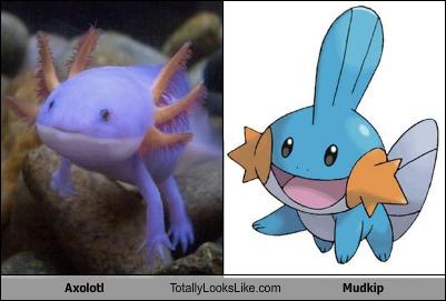
So i herd u liek mudkipz – Axolotl

Just as the “so i herd u liek mudkipz” meme has popularized the water type starter, Mudkip, from the beloved Ruby, Sapphire, and Emerald Pokémon games (and, recently Omega Ruby and Alpha Sapphire), the internet seems to have somewhat familiarized the public with a peculiar and endangered salamander.

Mudkip beside an axolotl Photoshopped to be the same colors.

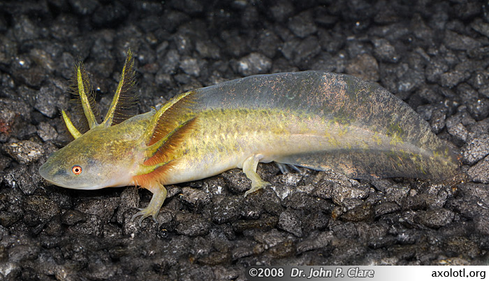
The Axolotl (Ambystoma mexicanum) lives exclusively in the fragmented remains of Central Mexico’s Lake Chalco and was once present in Lake Xochimilco before it was drained. These salamanders can come in a variety of different colors, two of them being wild type and the other two present in mutations. The wild colors are brown and black, while the mutant colors are leucistic and albino. Although both the leucistic and albino axolotls are varying shades of pinkish white, the albino has pink eyes and the leucistic has black eyes. They are closely related to tiger salamanders, sharing the same genus, and in fact look very similar to the larvae of tiger salamanders. Unlike other salamanders, axolotls normally never undergo metamorphosis into an adult form. This does not mean that they are not sexually mature, however. The average axolotl reaches maturity at 18-24 months of age with a typical length of around nine inches. They have both rudimentary lungs and gills so they are able to take oxygen from the water and gulp air at the surface. The gills are arranged on three stalks on each side behind the head. An axolotl also retains fins running from the end of its body to the tip of the tail.

Barred tiger salamander larva (Ambystoma mavortium mavortium).

4571101149_3b91879a14_c

Adult axolotl with wild type coloration

Retaining larval characteristics throughout life is called neoteny. Other salamanders can become neotinic if conditions in the environment put an unusual level of stress on the animal, but the axolotl is exclusively neotinic within its species. On extremely rare occasions, axolotls will progress to adult form. They most often remain strictly aquatic, though. They are naturally top predators in the areas they inhabit and feed on mollusks, crustaceans, water insects, and small fish. Since they are neotinic, their teeth are not fully developed and are only rough stumps in their jaws. They use these tooth stumps to grasp a prey item and position it before swallowing it whole. Unfortunately, since the introduction of large, nonnative fish, there is more competition for food and occasional predation upon axolotls by these fish. Additionally, axolotls are sold in markets as a delicacy which further reduces their population and they are now a red listed species that is critically endangered.

tumblr_o83gelQne01tic84no2_1280

Metamorphosed leucistic axolotl

Another very intriguing aspect of the axolotl is its Wolverine-like ability to heal itself. This trait has made it one of the most heavily studied salamanders in the world. Axolotls can completely regenerate their limbs, spinal cords, and even some organs in a matter of a couple months. The younger the salamander, the faster it heals. Older axolotls can still regenerate, but it takes much longer. The oldest axolotl in captivity, named Tiger Girl, in the Axolotl Center of Hanover, Germany was 16 years old (as of 2010) and it took her a year to grow back a leg that was bitten off by one of her tank mates. This research center is dedicated to the conservation of axolotls and studies their regeneration abilities in a humane, minimum stress environment. Researchers will anesthetize a salamander and cut off part of its limb to study the healing process. This doesn’t hurt the animal due to its incredible healing abilities and it is well cared for and eventually left to a life of retirement with no more testing.

Video about the Axolotl Center:
http://www.spiegel.de/international/zeitgeist/miracle-healer-scientists-attempt-to-crack-secret-code-of-the-axolotl-a-732283.html

Research institutions like the one in Hanover are helping to strengthen the international breeding program of axolotls and conserve this very remarkable and interesting species. It may give us clues about how to help humans who are injured to heal more effectively, such as burn victims. It is not only a fascinating animal, but also a valuable source of information for science and medicine that is imperative to save.

References:
1. Luis Zambrano et al. “Ambystoma mexicanum”. The IUCN Red List of Threatened Species, 2015. Web. 23 June, 2015. http://www.iucnredlist.org/details/1095/0

2. Taurog, Alvin, et al. “The role of TRH in the neoteny of the Mexican axolotl (Ambystoma mexicanum).” General and comparative endocrinology 24.3 (1974): 267-279.

3. Kragl, Martin, et al. “Cells keep a memory of their tissue origin during axolotl limb regeneration.” Nature 460.7251 (2009): 60-65.

4. Hamm, Magdalena. “Miracle Healer.” Speigel Online International. Speigel, 2 Dec. 2010. Web. 23 June 2015. <http://www.spiegel.de/international/zeitgeist/miracle-healer-scientists-attempt-to-crack-secret-code-of-the-axolotl-a-732283.html&gt;.

Photo and Video Links:
1.https://creaturefacts.org/wp-content/uploads/2015/07/afa4f-axolotl-totally-looks-like-mudkip.jpg

2.http://www.axolotl.org/images/tiger/tiger-salamander-larva.jpg

3.https://flic.kr/p/7XW67M

4.https://alphynix.tumblr.com/post/145316913155/edge-of-existence-edge-it-is-oddly-enough/embed

5.http://www.spiegel.de/international/zeitgeist/miracle-healer-scientists-attempt-to-crack-secret-code-of-the-axolotl-a-732283.html

6.http://i.dailymail.co.uk/i/pix/2013/05/17/article-2326057-19D49A37000005DC-864_634x419.jpg

Tiny King of the Desert – Southern Grasshopper Mouse

True or False:
All mice only eat seeds and other plant based food.

If you chose true, you’re wrong!

There is actually such thing as a carnivorous mouse! It lives in the Sonoran Desert of the American Southwest and Northern Mexico and is the worst nightmare of many an arthropod. The Southern grasshopper mouse (Onychomys torridus) is a tiny and ridiculously vicious predator of anything from deadly Arizona bark scorpions (Centruroides sculpturatus), tarantulas, giant desert centipedes (Scolopendra heros), and lizards, to even other small mammals – its own species included. Larger prey items like lizards or rodents are killed with a bite through the spinal cord at the base of the skull. Other prey are ripped apart and eaten bit by bit.

Being mice, these animals are small, but they are able to take down prey much larger than themselves. They reach maturity at a young age, as do other mice, and are weaned of their mother’s milk and able to eat whatever is brought to them by 20 days. Litters consist of 1-5 mice and these babies become sexually mature by day 40. Females have their first litters between 4 and 5 months of age and raise them in a burrow stolen from another rodent (which may have been killed and eaten during the eviction process).

grasshopper_mouse_scorpion

One of the most impressive things about Southern grasshopper mice is that they are able to take on venomous scorpions like C. sculpturatus with no ill effects. How do they do this?

When it comes to scorpion stings, these mice just don’t care. They don’t just ignore the pain, they don’t even feel it. This is because they have special pain pathways that are affected very differently by scorpion venom than other mammals. Normally, painful sensations are sent to the central nervous system (CNS) via the tetrodotoxin-sensitive (TTX-S) Nav1.7 and tetrodotoxin-resistant (TTX-R) Nav1.8 voltage-gated sodium channels in the dorsal root ganglia (DRG). The Nav1.7 is activated while Nav1.8 is left alone, and a signal is sent to the CNS.
Ashlee Rowe and colleagues experimented in the lab to find out how the venom affects these sodium channels in the grasshopper mouse. They injected nonlethal doses of Centruroides venom into the paws of both grasshopper mice and house mice (Mus musculus), which served as the control species. In addition to venom, they also injected saline and formalin, another painful chemical. These served as negative and positive controls, respectively. The behavior of the mice and the responses of the DRG neurons were measured.
As expected, house mice spent a lot of time licking the paws injected with venom and with formalin, but showed little response to saline. In contrast, grasshopper mice with venom injections only licked their paw for a few seconds before returning to normal behavior. They actually showed more irritation in response to the saline than the venom. When treated with formalin, the grasshopper mice licked the injured paw significantly more than in response to venom, but still less than house mice did. This indicates that their resistance to pain is specific to scorpion venom, and does not spread to other pain inducing chemicals.

When sodium currents in the Nav1.8 DRG neurons were measured in grasshopper mice, it was found that the venom actually inhibited these currents on a dose dependent scale. House mouse neurons showed no alteration in current, as is typical for most mammals. For Nav1.7 action potentials to be sustained after initial propagation, the sodium currents in Nav1.8 are necessary. Under the influence of scorpion venom, the researchers predicted inhibition of Nav1.8 to decrease excitation of the membranes and firing of action potentials. This is exactly what they discovered. The venom significantly decreased the excitation and blocked firing in grasshopper mouse neurons. The opposite occurred in house mice, supporting the expected, standard mammal response.
In short, the grasshopper mouse feels no pain in response to Centruroides venom thanks to the signals never reaching the CNS. Understanding how this mouse’s nociceptors work differently could have implications in medical treatment for human pain.
Whew! That was a lot of complicated science! Let’s get back into something a little more fun and simple.
Can this little rodent get any more badass than it already is? The answer is yes, it can! Not only do grasshopper mice take down and devour some of the most fearsome creatures in the desert, they also advertise their reign as tiny kings with howling war cries. That’s right! These mice howl at the moon like miniature wolves. As a species, they are very territorial and will let others know who the top mouse is by rearing up on their hind legs, throwing back their heads, and emitting a high, carrying howl after they make a kill.

If you ever go camping in the Sonoran desert and you hear the strange, whistling cry of a grasshopper mouse, you can be pretty sure that there’s one less bark scorpion to come crawling into your tent at night.

References:
1. McCarty, Richard. “Onychomys torridus.” Mammalian Species (1975): 1-5.

2. Reardon, Sara. “Mouse Eats Scorpions and Howls at the Moon.” Zoologger. New Scientist, 11 Jan. 2013. Web. 14 July 2015. <https://www.newscientist.com/article/dn23072-zoologger-mouse-eats-scorpions-and-howls-at-the-moon/&gt;.

3. Wade, Lizzie. “Mouse Impervious to Scorpion’s Sting.” Science Magazine. AAAS, 24 Oct. 2013. Web. 14 July 2015. <http%3A%2F%2Fnews.sciencemag.org%2Fbiology%2F2013%2F10%2Fmouse-impervious-scorpions-sting>.

4. Rowe, Ashlee H., et al. “Voltage-gated sodium channel in grasshopper mice defends against bark scorpion toxin.” Science 342.6157 (2013): 441-446.

Photo and Video Links:
1. https://www.youtube.com/watch?v=AcYW31N96b4

2.http://phenomena.nationalgeographic.com/files/2013/10/Grasshopper_mouse_scorpion.jpg

3.http://www.mammalogy.org/uploads/imagecache/library_image/library/1170.jpg

4. https://www.youtube.com/watch?v=9Xa-GSm1P0I

There’s Nothing Funny About These – Stinkhorns

phallus-hadriani-624d8644-05ea-49a1-b010-7f39a1c6b03-resize-750
What? Why are you laughing? Get your mind out of the gutter! Jeez!

Stinkhorns are not at all hilarious looking mushrooms from the fungus family, Phallaceae. They are found all over the world, though more so in tropical environments. They start out as what are called “eggs” under the ground. As they grow, these eggs rise to the surface and develop into their fruiting body form within just a few hours. This fruiting body is the mushroom that is visible to us on the surface and is used for reproduction. There are many different species of stinkhorns and they come in a multitude of differing shapes, colors, and sizes. Most have long, whitish and spongy shafts with a brown to puke green cap or “head”. Some, like the “ornate stinkhorns”, can look like anything from a beached octopus to a children’s climbing dome from hell. The egg-like start to the fruiting body is shared by all these species as is a gross looking spore slime, called gleba, that smells strongly of excrement and attracts insects to carry away the spores.

Mutinus ravenellii (Credit: Roberto Zanon)

Stinkhorns are not particular about where they pop out of the ground, and will often show up in people’s gardens, lawns, or other places where ripe smells aren’t generally appreciated. Most species fruit in summer and only last for a short period of time, however. Despite this, there are many internet articles on how to get rid of and prevent stinkhorn invasions. Stinkhorns can actually be beneficial to gardeners because they break down dead matter into nutrients that sustain plant life, and are self-sufficient. If only they didn’t smell so awful.
Strange as these fungi are, some people still consider them to be a delicacy. One stinkhorn in particular, the long net stinkhorn (Phallus indusiatus), is especially coveted in parts of China where it is eaten in egg form, dried mushroom form, pickled, stewed, and many other ways. This species tries to be a little fancier than its relatives by wearing a lacy skirt.

To the American palate, P. industiatus is described as “markedly unpleasant” in texture and bland in flavor. Personally, I’m not a huge fan of eating mushrooms to begin with, let alone one that is naturally covered in goopey, butt scented spore slime. But hey, if that’s what you’re into, then more power to you. Just don’t mistake the stinkhorn egg stage for the eggs of the poisonous Amanita mushroom.

Penis.

References:
1. Kuo, M. “Stinkhorns: Phallaceae and Clathraceae”. MushroomExpert.Com. Mushroom Expert, April 2011. Web. 10 July 2015. http://www.mushroomexpert.com/phallaceae.html

2. Williamson, Joey. “Stinkhorn Fungi – What Is That Smell?” Clemson Cooperative Extension. Clemson University, Nov. 2008. Web. 10 July 2015. <http://www.clemson.edu/extension/hgic/hot_topics/2008/11stinkhorns.html&gt;.

3. Hodge, Kathie T. “An Unlikely Delicacy: The Basket Stinkhorn :.” Cornell Mushroom Blog. Cornell University, 18 Feb. 2015. Web. 10 July 2015. <https://blog.mycology.cornell.edu/2015/02/18/an-unlikely-delicacy-the-basket-stinkhorn/&gt;.

4. Armstrong, W. P. “The Amazing Kingdom of Fungi.” The Amazing Fungi. Polamer College, 25 Jan. 2010. Web. 10 July 2015. <http://waynesword.palomar.edu/ww0504.htm&gt;.

Photo Links:
1.https://alchetron.com/Phallus-hadriani

2.https://upload.wikimedia.org/wikipedia/commons/thumb/3/3e/Mutinus_ravenelii_01.jpg/220px-Mutinus_ravenelii_01.jpg

3.https://upload.wikimedia.org/wikipedia/commons/b/b7/Phallus_indusiatus_02.JPG

4.http://31.media.tumblr.com/06eb892cc3fa8b494f8a4a521ad94c2f/tumblr_mwoq5otGWw1r3clqao1_500.gif

Extreme Dedication – Darwin’s Frog

This cute little frog might not look extraordinary in any particular way, but it has one of the strangest reproductive behaviors in the world. The Darwin’s frog exists as two separate species: The Chile Darwin’s frog (Rhinoderma rufum) and the Southern Darwin’s frog (R. darwinii). Like many other frogs, Darwin’s frogs live in the damp litter of the temperate forest floor within their habitat range and feed on small insects. The frogs’ backs vary greatly in coloring and come in pale green, dark green, tan, reddish brown, dark brown, and several other different shades. All of them have cream colored bellies with black blotches of differing size. Chile Darwin’s frogs are found only in Chile, as the name suggests, from Curico to Araouco Province. The Southern Darwin’s frog occurs in central and south Chile as well as nearby areas of Argentina. Both species are on the decline, but the Chile Darwin’s frog is more so and is listed as a critically endangered species in the IUCN Red List of Threatened Species. It may already be extinct, as the last recorded sighting was in 1980. Reasons for the decline of these frogs ranges from habitat loss to a devastating fungal disease, chytridiomycosis (caused by the Batrachochytrium dendrobatidis fungus), that is particularly destructive to amphibians.

5414255485_5b08fa7368_c

A frog killed by chytridiomycosis. Taken by Brian Gratwicke on Flickr.

12248364886_047e93d28e_c

2071602239_1518e8058f_c

Two different color morphs of R. darwinii: First photo by Flavio Camus and second by Efe, both from Flickr.

What makes these two frog species so unusual and unique among animals is the fact that the tadpoles develop inside the male frog’s vocal sac. After the female lays her eggs, the male will guard them until they hatch, about eight days. At this point, he takes them into his mouth and they go into his vocal sac through a slit where they stay to develop. A male Darwin frog’s vocal sac is much more expanded than a normal frog’s and runs the length of his belly. It also produces special secretions that provide the young with nutrients. The only difference between species when it comes to the technique involved with this brooding method is time. The Chile Darwin’s frog male only keeps the tadpoles in his vocal sac for the first few stages of development, then takes them to a nearby stream or pool and releases them to mature fully. The Southern Darwin’s frog actually carries the tadpoles full term. These tadpoles may remain in his vocal sac for up to 50 days. When they are totally developed, he then regurgitates them as tiny froglets.

It is not yet known how or why this weird brooding behavior evolved and this is still a topic of research. One reason may be that it eliminates the dangerous period of time spent as a tadpole under the constant watch of predators. Most tadpoles of other frogs don’t survive to adulthood, but the Darwin’s frog tadpole receives the ultimate protection while in a male’s vocal sac. As strange a method as it may be, this form of reproduction has worked out well for these two species of Darwin’s frog and, with continued research and conservation, it will hopefully work for them for years to come.

References:
1. Bourke, J., et al. “Batrachochytrium dendrobatidis in Darwin’s frog Rhinoderma spp. in Chile.” Diseases of aquatic organisms 92.2 (2010): 217.

2. Soto-Azat, Claudio, et al. “The population decline and extinction of Darwin’s frogs.” (2013): e66957.

3. “Darwin’s Frog (Rhinoderma darwinii).” Arkive. Wildscreen Arkive, n.d. Web. 24 June 2015. <http://www.arkive.org/darwins-frog/rhinoderma-darwinii/&gt;.

4. Alberto Veloso et al. Rhinoderma rufum. Red List. The IUCN Red List of Threatened Species, 2010. Web. 24 June 2015. < http://www.iucnredlist.org/details/19514/0&gt;

5. Whittaker, Kellie and Chantasirisivisal, Peera. Rhinoderma rufum. AmphibeaWeb. 16 Feb. 2006. Web. 24 June 2015. < http://amphibiaweb.org/cgi-bin/amphib_query?where-genus=Rhinoderma&where-species=rufum&gt;

Photo and Video Links:
1. https://flickr.com/photos/briangratwicke/

2. https://flickr.com/photos/chicuco/

3. https://flickr.com/photos/huenchecal/

4. https://www.youtube.com/watch?v=IAF5N-HwgOc

The Real Life ‘Alien’ – Cotesia congregata

I’m sure, even if you haven’t seen them, most of you are at least somewhat familiar with the movies in the Alien franchise and the premise behind them. You’re probably extremely grateful that there aren’t any terrifying creatures like xenomorphs stalking the earth, right? Well, that’s not entirely true. For tobacco hornworms (Manduca sexta), the caterpillars of a species of sphinx moth throughout the Americas, the horrifying scenario of Alien is a regular occurrence. This is thanks to a tiny parasitoid wasp called Cotesia congregata in the braconid family. Parasitoids are different from parasites in that they depend on the host for survival and keep it alive until they no longer have a use for it. At this point, they move on to a different stage of life, destroying the host in the process.

When it comes time to reproduce, the female wasp seeks out a larval tobacco hornworm and lays her eggs inside the body of the unfortunate caterpillar. With the eggs, she deposits a special symbiotic polyDNAvirus as well. This virus lives and multiplies exclusively inside the wasp’s reproductive system and is like a part of the wasp itself. The virus can’t reproduce on its own because it doesn’t possess the necessary genes to replicate. Instead, the genetic codes for the virus are present in the wasp’s genome. When this virus enters the caterpillar along with the eggs, it infects the caterpillar’s cells and dismantles its immune system. This protects the wasp eggs from being encapsulated and killed by the caterpillar’s hemocytes (insect blood cells). In addition to immune suppression, the virus arrests the development of the host and prevents metamorphosis into a moth. It also causes the accumulation of certain proteins in the host’s nervous system that allow it to survive for longer periods of time without food or water and alters its metabolism. This serves to keep the host alive to protect the developing wasps until they move onto their next life stage.

A fantastic close-up by Doug Smith on bugguide.net.

While inside the tobacco hornworm caterpillar, the C. congregata larvae hatch from their eggs and feed on the host while they grow larger and larger. By the time they are old enough to move on, the larvae have essentially eaten the host from the inside and chew their way out of its body to spin cocoons so they can pupate into adults. The larvae don’t travel away from the caterpillar at all to do this and create their cocoons right there on its body. After they pupate, the wasps emerge as fully formed adults and take to the air in search of mates in order to start the gruesome cycle all over again.

This poor fellow will never fulfill its destiny to become a moth

As terrible as it is for the hosts, C. congregata and many other species of parasitoid wasps are used as a means of biological control for a myriad of Lepidopterans (butterflies and moths) and other insects. There are many different species of parasitoid wasps and each of them primarily parasitizes a specific host insect. Many hosts are considered agricultural pests and wasps can be purchased and released to do their horrifying work without the destructive effects of pesticides or other artificial pest control techniques. Still, not all the insects that parasitoid wasps prey upon become hosts. The majority of them get to develop into their adult stage like our intriguing friend, the tobacco hornworm.

This one made it to adulthood! Congratulations on not dying!

References:
1. Shaw, M. R., and T. Huddleston. “Classification and biology of braconid wasps.” Handbooks for the identification of British insects 7.11 (1991): 126.

2. Beckage, Nancy E., et al. “Characterization and biological effects of Cotesia congregata polydnavirus on host larvae of the tobacco hornworm, Manduca sexta.” Archives of Insect Biochemistry and Physiology 26.2‐3 (1994): 165-195.

3. Beckage, Nancy E., and Michael R. Kanost. “Effects of parasitism by the braconid wasp Cotesia congregata on host hemolymph proteins of the tobacco hornworm, Manduca sexta.” Insect biochemistry and molecular biology 23.5 (1993): 643-653.

4. Beckage, Nancy E., and Dale B. Gelman. “WASP PARASITOID DISRUPTION OF HOST DEVELOPMENT: Implications for New Biologically Based Strategies for Insect Control*.” Annual Reviews in Entomology 49.1 (2004): 299-330.

Photo Links:
1.http://www.mdpi.com/insects/insects-03-00091/article_deploy/html/images/insects-03-00091-g001-1024.png

2.https://bugguide.net/node/view/76965

3.https://commons.wikimedia.org/wiki/File:Caterpillar_with_eggs1.jpg

4.http://www.nsf.gov/news/mmg/media/images/Manduca_sexta13b_h.jpg

Jaws Full of Teeth – American Crocodile

Chances are, when someone mentions crocodiles, you most likely immediately think of the Nile and saltwater crocs of Africa and Australia, respectively. The fact is, these ancient monster reptiles are so evolutionarily successful that they occur in the Americas too. The American crocodile (Crocodylus acutus) is found throughout the waters along the Pacific coast of Mexico and Central and South American down to Ecuador. It is even present on the Southern tip of Florida in the United States and in the Caribbean islands. The highest concentration of American crocodiles is in Lago Enriquillo, a hypersaline lake in the Dominican Republic. It’s not surprising how widespread crocs are given that they’ve been on the earth for 84 million years – even before dinosaurs.

Sarcosuchus imperator skull compared with extant crocodile species.

The American crocodile is one of the largest crocodilian species in the world and South American male crocs can reach lengths of up to 20 feet. Lengths of 14-16 feet are much more common, however, and U.S. crocodiles generally don’t exceed 13 feet. At sizes like these, they do pose a significant threat to humans and are more aggressive than American alligators. Although very rare, human killings have been recorded. American crocs are very tolerant of saltwater, hence their other common name of American saltwater crocodile, and possess special glands in their tongues that secrete excess salt to keep their internal saline concentration in balance.

Crocodiles are air breathing creatures and are therefore buoyant to a certain degree due to the air in their lungs when they inhale just as any other land animal. Unfortunately, they wouldn’t be able to hunt if they were just floating on the surface of the water all the time, or even slightly submerged. Their prey would see them for sure. But being the reptilian masterminds that they are, they’ve developed a strategy to combat this. Crocodiles will seek out stones and swallow them to create a ballast. This way, they can sink themselves to the bottom or rise up to the surface whenever they please. Mostly they submerge everything but their eyes and nostrils and lie in wait for anything edible to wander too close, but they will also hang out on the bottom of a river or lake too and are capable of staying under for hours. American crocodiles eat anything from large mammals and birds to crabs, fish, and snails. They will also sometimes scavenge for carrion.

24590885137_5d37401ae6_z

An American crocodile in Florida, that seems to have an old jaw injury, eats a fish. (By Andrew DuBois on Flickr)

When it comes to breeding, usually in late fall or winter, American crocodiles will come together and partake in surprisingly gentle and elegant courtship that can last up to a month before any mating takes place. The female will then excavate a burrow, or if no burrowing sites are available, create a mound in which to lay her eggs. The material for the nest and amount of incubation is extremely important because sex is determined by temperature and a subtle fluctuation out of optimal range could create a clutch of either all female or all male crocodiles. This would decrease genetic variation in the species and make them more vulnerable to disease and other factors that can devastate populations. Both parents stay and aggressively protect the nest until the babies hatch, then the female provides them with protection and transportation for several weeks until they disperse.

16204000462_89a1331dfd_z

A baby American crocodile by “ryanacandee” on Flickr.

As big and heavy as American crocs are and as lazy and lethargic as they appear to be most of the time, their agility is not to be underestimated. These huge reptiles can swim up to 20 miles an hour and run on land at 10. When they are walking, trotting, or running, they move their legs underneath and raise their bodies off the ground. Freshwater crocodiles can actually gallop and reach speeds of 15 miles per hour.

This is a Nile crocodile, but it gives you an idea of what they look like when they run.

Here’s a video of a freshwater crocodile galloping as well:

So if you ever see a wild crocodilian of any kind, don’t assume you can just run away from it if you get too close. Stand way, way back and enjoy one of the most perfect, unchanged predators on earth.

In memory of Lolong, the world’s largest captive crocodile, who died on February 10th, 2013 in a conservation center in the Philippines. He was a 20.3 foot long saltwater crocodile and is thought to have been over 50 years old. His cause of death is still unknown.

References:
1. Fishman, Jake and MacKinnon, Kristin. “Crocodylus acutus (American Crocodile)”. Animal Diversity Web. University of Michigan Museum of Zoology, 2009. Web. 24 June 2015. http://animaldiversity.org/accounts/Crocodylus_acutus/

2. “American Crocodiles.” National Geographic. National Geographic Society, n.d. Web. 24 June 2015. <http://animals.nationalgeographic.com/animals/reptiles/american-crocodile/&gt;

3. Swiman, Elizabeth, Mark Hostetler, and Sarah Webb Miller. “Living with Alligators: A Florida Reality.” EDIS New Publications RSS. University of Florida, Aug. 2005. Web. 24 June 2015. <http://edis.ifas.ufl.edu/uw230&gt;.

4. Taplin, Laurence E., et al. “Lingual salt glands in Crocodylus acutus and C. johnstoni and their absence from Alligator mississipiensis and Caiman crocodilus.” Journal of Comparative Physiology B: Biochemical, Systemic, and Environmental Physiology 149.1 (1982): 43-47.

5. Wedel, Matt. “Eusuchia.” Unversity of California Museum of Paleontology. University of California, Berkely, May 2010. Web. 24 June 2015. <http://www.ucmp.berkeley.edu/taxa/verts/archosaurs/eusuchia.php&gt;.

6. Parrish, J. Michael. “The origin of crocodilian locomotion.” Paleobiology (1987): 396-414.

7. Britton, Adam. “Lolong Officially the World’s Largest Crocodile in Captivity.” Weblog post. Croc Blog. Crocodilian.com, 23 June 2012. Web. 24 June 2015. <http://crocodilian.blogspot.com/2012/06/lolong-officially-worlds-largest.html&gt;.

Photo and Video Links:
1.http://img3.wikia.nocookie.net/__cb20120817051721/dinosaurs/images/7/79/Sarcosuchus.jpg

2.https://flic.kr/p/3AqRmw

3.https://flic.kr/p/Dt1Hqe

4.https://flic.kr/p/qFTJNj

5.http://25.media.tumblr.com/tumblr_m6s3ch2JN81r3zfc1o1_500.jpg

6.https://www.youtube.com/watch?v=d_Qmi8MP5uI

The Cock-Blocking Flower – Bee Orchid

Bee orchids of the genus Ophrys are such seductive flowers to male bees and wasps that the insects actually prefer the sexy smell of the orchids to the scent of ready, virgin female bees and wasps. These many species of flowers are found in central and South Europe, North Africa, Asia Minor, and the Caucasus Mountains. Most of them are from around the Mediterranean, however. Once an orchid grows in its spot, it is very difficult to transplant because of the symbiotic fungi in its roots that it needs in order to live. Disturbing this relationship usually ends badly for the orchid, so it is not easy to cultivate. The leaves are at the base of the plant and have a greenish blue coloring with a very shiny surface. The flowers of different species vary as much as the insects they attract. Each distinct species relies solely on a specific insect for its pollination. The types of insects that pollinate bee orchids are typically bees and wasps, but some varieties depend on beetles.

Ophrys apifera

Ophrys lupercalis with male bee

Everything about a bee orchid flower is designed to lure and seduce horny male insects to come and make love to it. It uses special chemicals to replicate the scent of a virgin female almost perfectly, but with slight differences. These small variations in scent are actually what make it so irresistible. Discrepancies in perfume signify differences in genetics and bees are naturally attracted to slight difference because it helps to prevent inbreeding. Ophrys blossoms also use tactile and visual mimicry to appeal to the sex-hungry boys. The lower half of the flower has the same shape and coloring of females of the target species and resembles a female visiting or resting on the flower. Velvety fuzz gives it the feel of a female and draws the bewitched male even deeper into the flower where it proceeds to mate with it and become coated in pollen. Once the insect figures out that it’s been tricked, the orchid has already done what it needed to do. When the stupid, love-struck bee, wasp, or beetle flies over and gets fooled by another orchid flower, pollination occurs and the whole thing starts over again.

50743750167_deb416d799_c

Ophrys bombyliflora by Adrien Chateignier on Flickr

Eventually, insects will catch on to the orchid’s sexual game and stop visiting those flowers altogether. Fortunately for the orchid, there are plenty more males that will fall for the disguise and they also produce up to 12,000 tiny seeds to go forth and create new plants, thus spreading their beautiful sexual deception across the land.

Ophrys insectifera with male wasp

References:
1. Govaerts, G. “ Ophrys L., Sp. Pl.: 948 (1753).” World Checklist of Selected Plant Families. Board of Trustees of the Royal Botanic Gardens, Kew, n.d. Web. 23 June, 2015. http://apps.kew.org/wcsp/namedetail.do?name_id=140696

2. Pollan, Michael. “Love and Lies.” National Geographic Magazine. National Geographic Society, Sept. 2009. Web. 23 June 2015. <http://ngm.nationalgeographic.com/print/2009/09/orchids/pollan-text&gt;.

3. Curtis, John T. “The relation of specificity of orchid mycorrhizal fungi to the problem of symbiosis.” American Journal of Botany (1939): 390-399.

Photo Links:
1.https://upload.wikimedia.org/wikipedia/commons/0/02/Ophrys_apifera.jpg

2.http://newsimg.bbc.co.uk/media/images/47682000/jpg/_47682074_lupercalis_466.jpg

3.https://flickr.com/photos/adrien2008/

4.http://www.orchid-nord.com/orchids_nord/ophrys_insectifera/Ophrys-insectifera-&-pollin.jpg

Nature’s Moste Potente Potion – Golden Poison Frog

For those of you wondering about the title, no it is not a typo. It is an obscure Harry Potter reference, and if you got it, I applaud you.
Remember that golden poison frog (Phyllobates terribilis) I mentioned in the last article about the hooded pitohui? Well, I thought it should get its very own entry because it’s just such a cool little thing. As I mentioned before, it is one of the few animals that carries the deadly batrachotoxin. It gets this poison from the beetles and ants it eats just, like the pitohui does. The frog, however, is much, much, much more dangerous! You can even get this from its Latin name – “terribilis” speaks for itself. So before I explain how toxic it is, let’s find out where they live and what they’re like so if you ever travel there, you can keep an eye out and avoid them like the plague.
The golden poison frog is found exclusively in the rain forests of Colombia in South America. Instead of living on the forest floor like many other poison frogs, it lives in the trees up to about eight feet above the ground. These poison frogs do come down occasionally, especially when feeding, but generally stick to high up plants. They don’t use bromeliad pools to lay their eggs, so they need to come down to the ground in order to reproduce as well. When the eggs hatch into tadpoles, the parents transport them to water filled tree hollows where they can metamorphose. Golden poison frogs, like other poison frogs, are very social animals and live in groups with up to several individuals in captivity.

3624354133_d52a5e4617_z

Now the part about their poison. Like I said, it is much stronger than the pitohui’s. It is a very important killing tool to the native people of Colombia when they hunt. Pinning a frog with a stick and rubbing a heated dart across its back can keep the dart effective for two years afterward. Some tribes even keep several of these frogs in woven baskets for their poison, but they must take great care in doing so. Inevitably, there are cases where people have died from accidentally touching a captured frog. The average frog contains around one milligram of poison, which is enough to kill up to 20 adult humans or two male African elephants. This can be translated to 15,000 humans per gram of poison. Not only is the poison toxic while on the frog itself, but it can last once rubbed off as well, as in the case of the poison darts. It is absorbed through the skin, so simply brushing against a frog or something its skin has touched can kill. Batrachotoxin attacks sodium ion channels on cells within the central nervous system, which causes paralysis of pretty much everything including the heart and lungs. This can lead to death for a person in five minutes. The golden poison frog has special sodium channels that aren’t affected by batrachotoxin, which is why it can carry such a deadly substance around without any harm to itself.

Ball and stick model of batrachotoxin’s molecular structure.

Funnily, these frogs are often kept by amphibian hobbyists as pets. It’s okay, though, because captive raised frogs aren’t toxic at all and can be handled freely for as long as the animal will tolerate it. All the poison comes from their diet and captive frogs’ diets are devoid of the prey that gives them their deadly defense. However, wild caught golden poison frogs are dangerous. Even after a change of diet, the toxins can linger in the frog’s body for years afterward, so it is inadvisable to keep anything but a captive bred golden frog. As the name states, they obviously come in a lovely, bright gold color, but there are also a variety of other color morphs. These include the orange morph and mint morph.

Mint morph

31647336532_65400c767d_z

Orange morph by John Clare on Flickr.

As beautiful as golden poison frogs are, stay well away if you ever go to Colombia and find one! You wouldn’t even be able to be sorry if you got too close because you’d be dead.

References:
1. Gratwicke, Brian. “Golden Poison Frog.” WAZA. World Association of Zoos and Aquariums, n.d. Web. 25 June 2015. <http://www.waza.org/en/zoo/choose-a-species/amphibians/frogs-and-toads/phyllobates-terribilis&gt;.

2. Patocka, Jiri, Kräuff S. Wulff, and María V. M. Palomeque. “Dart Poson Frogs and Thier Toxins.” Ed. Colonel R. Price. The ASA Newsletter 74 (1999): n. pag. Applied Science and Analysis. Web. 24 June 2015. <http://www.asanltr.com/ASANews-99/995frogs.htm&gt;.

3. Stuart, Sean K. “The True Poison-Dart Frog: The Golden Poison Frog.” Frogs. Herpetologic.net, n.d. Web. 24 June 2015. <http://www.herpetologic.net/frogs/caresheets/terribilis.html&gt;.

4. Alvarez, Mariela C. and Wiley, Mary. “Phyllobates terribilis”. Animal Diversity Web. University of Michigan Museum of Zoology, 20 Aug. 2011. Web. 24 June 2015. http://animaldiversity.org/accounts/Phyllobates_terribilis/

5. Wunderli, Michael. “Golden Poison Frog (Phyllobates Terribilis)” Encyclopedia of Life. Eol.org, n.d. Web. 25 June 2015. <http://eol.org/pages/1039238/details&gt;.

Photo Links:
1.https://flic.kr/p/6wgLdn

2.http://www.batrachotoxin.com/batrachotoxin720.gif

3.http://vignette4.wikia.nocookie.net/reptiles/images/4/44/Cuddling_Terribilis.jpg

4.https://flic.kr/p/QdySzj

The Poison Bird – Hooded Pitohui

The hooded hitohui (Pitohui dichrous) of Papua New Guinea is one of the world’s first and only documented poisonous birds. It was discovered to be toxic during a 1989 study of birds of paradise by American ornithologist John “Jack” Dumbacher. He and his team had mist nets set out to catch birds of paradise, but inevitably, other birds were caught in the nets as well. Some of these birds were hooded pitohuis, which would bite and scratch defensively when being extracted from the mist net. Dumbacher got a bite from one of the birds on his finger that started to sting, so he decided suck on the cut to clean it. After about a minute, his lips and tongue started to tingle and burn. This happened to several other volunteers as well so the team wondered if the pitohui was the cause. When more pitohuis were captured, Dumbacher took a feather and put it directly on his tongue, finding that the burning and tingling effects could last for hours. When he returned to America, Dumbacher took some feathers to chemist John Daly at the National Institute of Health. By 1992, Daly discovered that the feathers contained batrachotoxin. It is an extremely powerful steroidal alkaloid poison that is largely neurotoxic, and even relatively small doses can lead to paralysis of all muscles, cardiac arrest, and then death. This is the most toxic natural substance known and is also present in the secretions of several poison dart frogs of the genus Phyllobates (most famously, Phyllobates terribilis – the Golden Poison Frog).

Credit: Brendan Freeman (Wikimedia Commons)

At first it was not known how this bird acquired such a potent toxin, but locals of New Guinea were asked what they knew about hooded pitohuis and relayed their observations. Pitohuis often catch and eat small and colorful Choresine beetles from the Melyridae family. The same toxin has previously been found in these beetles, which are also known to be preyed upon by Phyllobates poison dart frogs. The striking orange and black feathers of the hooded pitohui are another indication of its toxicity. Many poisonous and venomous animals are brightly colored to warn potential predators that they are very dangerous and not to be messed with. This is called aposematic coloration. Any animal unlucky enough to catch a pitohui would quickly learn to stay away when it sees those colors again. Of course, the bird has not always had this defense. First it had to evolve to cope with the poison itself before being able to incorporate the toxin as part of its own defense. It is likely that the first pitohuis that fed on melyrid beetles only ate a few at a time, slowly building up a resistance to batrachotoxin over generations. This toxin has been found in not only the bird’s feathers, but also its flesh.

This amazing, colorful bird just goes to show that even the most seemingly harmless animals can have alarming tricks up their sleeves.

References:
1. Dumbacher, John P., et al. “Homobatrachotoxin in the genus Pitohui: chemical defense in birds?.” SCIENCE-NEW YORK THEN WASHINGTON- (1992): 799-799.

2. Dumbacher, John P., et al. “Melyrid beetles (Choresine): A putative source for the batrachotoxin alkaloids found in poison-dart frogs and toxic passerine birds.” Proceedings of the National Academy of Sciences of the United States of America 101.45 (2004): 15857-15860.

3. “Jack Dumbacher.” California Academy of Sciences. N.p., n.d. Web. 24 June 2015. <http://www.calacademy.org/explore-science/jack-dumbacher&gt;.

Photo Links:
1. https://commons.wikimedia.org/wiki/File:Hooded_Pitohui_3.jpg

Beautiful, but Dangerous – Blue-Ringed Octopus

Today I will talk about a fantastic little creature called the blue-ringed octopus (Hapalochlaena lunulata). This is yet another venomous animal that haunts the waters of Australia as well as the Pacific and Indian Oceans. There are several species within the Hapalachlaena genus and I will be writing about that, as all species share the same general characteristics.

Though it is very small and vulnerable, the blue-ringed octopus is fairly docile unless provoked. It likes to hunt in tide pools and shallow reef waters where it scrounges out small crabs and shrimp that are killed quickly with its powerful venom. It is diurnal, so it mainly only comes out to hunt during the daytime. When it catches a prey item such as a small crab, it uses its sharp beak to break open the shell and eat out the insides.

When disturbed, the blue-ringed octopus will show the elaborate coloration that it is named for. Chromatophores in cephalopods such as octopuses, squid, and cuttlefish, are units of neuron controlled glial, muscle, and sheath cells. These are contained in an elastic sac that can expand and contract to change the opacity and translucency of the pigment within. The blue-ringed octopus is unusual in that it does not have chromatophores over its rings, but only surrounding them and underneath them. The iridescent, electric blue pigments in its skin are always present, yet only visible when it wishes them to be. Certain muscles above the iridophores, specialized light reflecting organelles within the cells, hide the blue-green light reflectors until they are contracted and expose the bright color. This is a very quick and highly controlled method of display that is not seen in other species of octopus.

16166613286_d2a61eb63e_k

Rings barely visible. Photo by Klaus Stiefel

3-9-13-blue-ringed-octopus1

Threat display

A blue-ringed octopus may only be from five to eight inches in length, but it packs potent enough toxins to kill an adult human in a matter of minutes. It has special salivary glands behind its beak containing bacteria that produce the deadly cocktail it delivers. An envenomation from one of these octopuses starts out with nausea and quickly progresses to numbness, difficulty swallowing, visual abnormalities, and eventually motor and respiratory arrest. Because there is little to no effect on cardiac muscles, a victim’s heart may continue to beat until asphyxia sets in and the central nervous system is deprived of oxygen completely. The bite itself may be painless and show only vague evidence of wound infliction. What makes the venom so deadly is the tetrodotoxin (TTX). This is the same toxin found in pufferfish and is designed to block sodium channels on neuron cell membranes, which in turn causes total paralysis throughout most vital areas of the body (minus the circulatory system). There is currently no quick fix in existence for blue-ringed octopus envenomation, so treatment is limited to life support measures, such as artificial respiration, until the effects of the venom wear off. Sometimes victims may be fully conscious and aware of their surroundings, but unable to move or speak due to the paralysis. Those who survive the first twenty four hours will typically make a full recovery.

3165716233_db1e6d4c0f_h

DO NOT DO THIS! (Unless you are a properly trained biologist). Photo by Steve Childs

Blue-ringed octopuses might be scary, but they are tender love-makers just like we imagine ourselves to be. A male blue-ring will court the female of his choice by approaching her and gently caressing her with his modified arm used for delivering sperm. He will then pull her close to him by grasping her mantle and use this arm to deposit packets of sperm inside her mantle cavity again and again until she is decides she is done. Sometimes the male has to be removed by force because he wants to keep going. Male blue-rings aren’t picky about who they court and will often attempt to mount other males of their own species. However, these interactions are usually much shorter lived and result in both males withdrawing, possibly in embarrassment, without a struggle or exchange of sperm. A little while after a female has been fertilized, she lays a single clutch of about fifty eggs and incubates them with her arms for up to six months. As with all octopuses, she does not eat during this time and dies shortly after her babies emerge from their eggs.

Unfortunately for such a pretty and intelligent animal, the maximum lifespan for a blue-ringed octopus is only two years. But hopefully it is two years well spent hunting, fascinating curious humans, and making sweet, sweet octopus love.

References:
1. Mäthger, Lydia M., et al. “How does the blue-ringed octopus (Hapalochlaena lunulata) flash its blue rings?.” The Journal of experimental biology 215.21 (2012): 3752-3757.

2. Yotsu-Yamashita, Mari, Dietrich Mebs, and Wolfgang Flachsenberger. “Distribution of tetrodotoxin in the body of the blue-ringed octopus (Hapalochlaena maculosa).” Toxicon 49.3 (2007): 410-412.

3. “Australian Marine Envenomations.” Marine Envenomation. University of Sydney, n.d. Web. 23 June 2015. <http://sydney.edu.au/medicine/anaesthesia/resources/venom/marine_enven.html#bluering&gt;.

4. Cheng, Mary W., and Roy L. Caldwell. “Sex identification and mating in the blue-ringed octopus, Hapalochlaena lunulata.” Animal behaviour 60.1 (2000): 27-33.

Photo Links:
1.http://blennywatcher.com/wp-content/uploads/Blue-ring-catches-crab3-BlennyWatcher.com_.png

2.Concealed rings

3.https://thesciencevine.files.wordpress.com/2013/09/3-9-13-blue-ringed-octopus1.jpg

4.Don’t touch

5.http://www.uwphotographyguide.com/images/Articles/durand_blue-ring-octopus-1.jpg