Could “The Last of Us” Really Happen?

UPDATE 4/19/22: Newer research has suggested that the ants that are infected with O. unilateralis are not in fact controlled via manipulation of the brain. Rather, there seems to be a chemical means by which the fungus forces the ant to bite the leaf vein. The study found no evidence of fungal cells inside the actual brain. However, hyphae were abundant throughout the head of the ant and the rest of its body, including inside the muscle fibers themselves, creating a complex network. The exact mechanisms behind the behavioral manipulation are still not known and more studies need to be done. With that in mind, please know that I am discussing the structure and function of ant brains in this article, as it helps exemplify the differences between an insect and vertebrate brain for the purpose of speculation.

For those who don’t know, The Last of Us is a dark, yet beautifully crafted survival horror game that explores what the world would be like if the majority of the human population were infected by a mutated strain of Ophiocordyceps unilateralis. This video game is critically acclaimed for its storytelling, graphics, and gameplay. It is considered a masterpiece by most of its reviewers and by its players. From the little bit I have played, I would absolutely agree. There are few developers that are able to take a real world scientific event, such as an insect being manipulated by a parasitic fungus, and implement the core concept realistically in a video game. Naughty Dog, the developer of The Last of Us, does this fantastically well.

71-hlREKk6L._AC_SY741_

Joel and Ellie are the game’s core characters

Ophiocordyceps unilateralis is in fact a real fungus. This species (actually a whole complex of species referred to collectively as Ophiocordyceps unilateralis sensu lato, meaning “in a broad sense” – I will refer to this species complex just as O. unilateralis from here) specifically targets ants, generally of the Camponotus genus, and takes over their bodies in order to move them to a location with ideal conditions for its reproduction. One of the most interesting things about this fungus is how precisely it manipulates its host. A 2009 study by Andersen et al. in The American Naturalist found that ants infected with O. unilateralis almost always died gripping a vein on the underside of a leaf (98%) and that this location was significantly more towards the north-northwest side of the plant than any other direction. Camponotus ants were found almost exclusively at a height of 25 centimeters from the forest floor, while closely related Polyrhachis ants were seen at a mean height of 78.43 centimeters. This kind of behavioral control is known as an extended phenotype. The gene expression of a parasite within a host’s tissue causes aberrant host behavior that increases the parasite’s fitness. Keep in mind that the ant species in the study colonize the canopy, not the forest floor, so this is extremely complex behavioral manipulation. Under the fungus’s control, these infected ants are traveling very far from their initial location. Many infected ants will often die in areas where others already have because the conditions in these places are favorable for O. unilateralis. This leads to places, called graveyards, that have a high density of corpses. Any healthy ant happening to walk through such an area will likely become infected by the deadly spores.

An ant with its jaws locked in a “death grip”.

When O. unilateralis spores come into contact with an ant, they adhere to it and start to penetrate its cuticle. After this, fungal cells start to infest the ant’s entire body and begin to alter its behavior. The infected ant then leaves the colony and starts the trek down toward the forest floor. Once a perfect spot is reached, the ant will bite the vein of a leaf and die with its jaws locked in place. This is known as the “death grip”, and keeps the ant from falling off the leaf. Any ants that end up dying within the colony due to infection will be carried far away by other workers. Within a few days, the stroma, or stalk, of the fungus will erupt from the back of its host’s head and develop a stromal plate that releases spores when mature. By this point, most of the ant’s original tissue has been replaced by fungus.

Saggital section of a dead Camponotus ant filled with fungus from The American Natrualist – W = white, Bl = black, O = orange, and P = pareithecial plate, which is where the spores originate. (The life of a dead ant: the expression of an adaptive extended phenotype. Andersen et al. 2009)

The ants in the video below are not Camponotus or Polyrhachis ants, and the fungus is not O. unilateralis so the hosts show a different behavior. However, it still illustrates the process well.

What a horror it would be if humans were susceptible to such a fate! The question is, could this actually happen? Let’s consider the ant brain for a moment. It has all the general features of a typical insect brain and is very simple compared to a vertebrate brain. However, ants have more well developed areas associated with memory and learning. These “higher” functions are controlled by the mushroom bodies. Mushroom bodies are much larger in worker ants than they are in the male soldiers. Workers must navigate varying spacio-temporal environments using multiple cues and also remember where food is and where home is. For ants, workers are the most “intelligent”. Still, one lone worker is of no use. It takes the whole colony to have a properly functioning and flourishing nest. An entire colony of ants, which can be up to millions of individuals, does not even come close to matching the complexity of a single human brain. If a fungus were to develop an ability to infect and alter the behavior of humans, this would probably take a ridiculously long time to evolve. Human beings would most likely not even be around any longer. But, let’s just roll with it and say that this could happen. What would it be like and how does The Last of Us compare?

Simplified ant brain from Recognition of social identity in ants (Bos and d’Ettorre, 2012).

We’ll start with the mode of infection. In the video game, people seem to become infected by breathing in the spores of the mutant O. unilateralis strain. Those who wear a special breathing bask in spore filled areas do not become infected. This appears to check out. Human skin is likely to be much less susceptible to becoming invaded by spores, simply because it is so much thicker and has many more layers than insect cuticle. Other fungi are able to infect hosts via inhalation (like some molds), so it would be no different for a mutant fungus; the infection would just result in a different pathology. So getting infected by this fungus is not in question if you breathe the spores in, but you can also fall victim if there is an exchange of bodily fluids, such as from a bite. This is logical as well. If the fungus has infiltrated an infected person’s system as in ants, bodily fluids like saliva and blood may contain fungal cells. These could easily be passed on to a healthy human.

What about the behavioral and morphological alterations? In The Last of Us, any infected individual, even in the early stages, is being driven by the fungus’ gene expression and aims to spread the spores or cells to as many others as possible. There are four main stages of infection from this mutant O. unilateralis: Runners, Stalkers, Clickers, and Bloaters. All of these collectively are referred to as the Infected (noun) in the game. Individuals who have just been infected are classified as Runners. They still cling to a shred of their humanity, however small, and try to resist the fungus’ behavioral control. However, if they are startled or as the infection progresses, they will snap and attempt to attack anyone nearby who is healthy. Stalkers are fully controlled by O. unilateralis and start to show fungal growth on the face. This fungus strain attacks the eyes first, so they have poor vision and begin to exhibit primitive signs of echolocation. In Clickers, the face has been fully engulfed by fungal growth and rendered the host completely blind. These Infecteds use clicking and screeching to crudely survey their surroundings within a small radius. The growth on the head and face forms a roughly dish shaped plate that helps to direct the clicks. Bloaters are the final stage and can take 10-15 years to develop. The entire body of this Infected is now covered in fungus, some of it showing bioluminescence, and is therefore very physically hardy. Bloaters are also able to throw sacs of toxic powder both as a defense and an attack mechanism. Once the host finally dies, whatever tissue is left of the former person crumbles and is used by the parasite to complete its growth and release spores.

Infected_Final_Stage

The remaining tissues of the victim begin to crumble as the fungus spreads its spores.

Now to analyze the behavior of the Infected. All stages appear to seek out specific locations and stay there, unless stimulated to pass on the infection when a healthy individual is sensed nearby. This reflects the behavior of O. unilateralis controlled ants that travel until they find the optimal conditions for the fungus’s reproduction and then fix themselves to that spot. However, infected ants will leave the colony and do not show any aggressive behavior whatsoever toward other ants. Why would the Infected exhibit such an aggressive drive to bite or kill? We actually have a disease in existence that does just this. It’s called rabies virus. If rabies can infiltrate the brains of mammals and stimulate this type of behavior, it would not be a stretch to think that a mutant fungus could affect humans this way. Rabies in humans doesn’t present the same way that it does in other mammals; it resembles a bad flu, followed by agitated behavior and cognitive deficits, sometimes with hallucinations, delirium, and insomnia. Perhaps our mutant O. unilateralis would be able to incite the aggressive behavior seen in the games by affecting the brain in a similar way to rabies in non-human mammals. This could explain the Infecteds’ responses to healthy humans.

Clickers use crude echolocation to detect the presence of nearby humans and then go into a full sprint toward anyone they sense.

Runners display behavioral tendencies that would be expected. Since the fungus has only just started infiltrating the brain, it is not surprising that the victim would be scared and confused by what is happening. It makes sense that a person in this state would be very volatile and dangerous to approach. Stalkers tend to hide out of sight and only engage when someone lets their guard down. This is presumably because their vision is extremely poor and they must wait until the opportune moment in order to successfully attack. Clickers are harder to explain realistically. It would be highly unlikely that an Infected would suddenly develop the ability to navigate using clicks. This is a skill that must utilize learning and memory, parts of the brain no longer accessible to the victim. Very few humans are able to echolocate and those who are have refined their technique over years. Real world O. unilateralis is not able to direct ants to do things they are not already capable of; it simply takes advantage of abilities the insect already possesses. The sense that would be most beneficial to a Clicker is touch. Of course, sound is still an effective way of interacting with surroundings, but it would not be used for spatial navigation as in The Last of Us. This can be extrapolated to the Bloaters in the game as well, because they also use echolocation. All of these Infected will attack to kill. This is presumably because the fungus cannot kill immediately on its own, but can reach maturity faster in a dead body. So, when controlling a living human, O. unilateralis uses that person to its advantage by driving them to help speed up reproduction through the provision of corpses. If the host fails to make a kill, but manages to penetrate the skin of a healthy human, then the fungus still benefits because that person will become another Infected.

Bloaters take 10 to 15 years to develop and have hard fungal growth all over their bodies, giving them great resistance to blunt and penetrating forces.

Overall, the concept of The Last of Us is scientifically plausible, given enough time for O. unilateralis or a different species of Ophiocordyceps fungus to evolve the traits explored here. The intelligence and thought put into this game has given it so much more than most other video games can offer. These things make it different from your standard zombie apocalypse. It is scary because it feels like it could really happen, and this realness comes from its grounding in actual science. The infection seen in it is already a reality for many insects and other arthropods. They may not show the aggressiveness displayed by the Infected, but the behavioral manipulation seen in these animals is extremely effective at fulfilling the fungus’s needs. Maybe at some point in the future, the fictional scenario of The Last of Us could come to be. Who knows? The natural world is continually revealing so many things previously thought impossible.

References:
1. Gronenberg, Wulfila. “Structure and function of ant (Hymenoptera: Formicidae) brains: strength in numbers.” Myrmecological News 11 (2008): 25-36.

2. Evans, Harry C., Simon L. Elliot, and David P. Hughes. “Ophiocordyceps unilateralis: A keystone species for unraveling ecosystem functioning and biodiversity of fungi in tropical forests?.” Communicative & integrative biology 4.5 (2011): 598-602.

3. Hughes, David P., et al. “Behavioral mechanisms and morphological symptoms of zombie ants dying from fungal infection.” BMC ecology 11.1 (2011): 13.

4. Andersen, Sandra B., et al. “The life of a dead ant: the expression of an adaptive extended phenotype.” The American Naturalist 174.3 (2009): 424-433.

5. Pontoppidan, Maj-Britt, et al. “Graveyards on the move: the spatio-temporal distribution of dead Ophiocordyceps-infected ants.” PloS one 4.3 (2009): e4835.

6. “The Infected.” The Last of Us Wiki. Wikia, 2013. Web. 7 Oct. 2015. <http://thelastofus.wikia.com/wiki/The_Infected>.

7. “Signs and Symptoms – Rabies.” Centers for Disease Control and Prevention. Centers for Disease Control and Prevention, 15 Feb. 2012. Web. 08 Oct. 2015.

Photo and Video Links:
1.http://static.giantbomb.com/uploads/scale_large/9/93770/2419553-397060_386577108098888_427807760_n.jpg

2.https://i.guim.co.uk/img/static/sys-images/Guardian/Pix/pictures/2010/12/21/1292935214310/a-dead-worker-ant-004.jpg

3.http://ento.psu.edu/publications/DH1

4.https://www.youtube.com/watch?v=XuKjBIBBAL8

5.http://journal.frontiersin.org/article/10.3389/fpsyg.2012.00083/full

6.https://vignette.wikia.nocookie.net/thelastofus/images/3/3f/Infected_Final_Stage.jpg/revision/latest?cb=20130224030859&format=original

7.http://vignette4.wikia.nocookie.net/thelastofus/images/9/9d/Clicker.jpg/revision/latest?cb=20150928101722

8.http://vignette4.wikia.nocookie.net/thelastofus/images/8/8f/Bloater.jpg/revision/latest?cb=20150928101859

There Will Be Blood – Stephen’s Banded Snake

It has already been established numerous times, but I feel like I should re-emphasize this well-known fact: Australia is a terrifying continent! The Stephen’s banded snake (Hoplocephalus stephensii) just gives us one more reason never to set foot in this country, specifically in New South Wales. H. stephensii has a feisty temperament and will readily bite those who try to handle it or accidentally threaten it in some way. As the “banded” part of its name suggests, this snake is covered in striking dark grey to black bands over and white or light brown body. It is a member of Elapidae, a family of snakes with hollow, usually small, fixed fangs (meaning they do not fold back when the mouth is closed as in vipers). The snakes in this family include cobras, taipans, sea snakes, brown snakes, tiger snakes, mambas, coral snakes, and many others.

stephens-banded-snake

Because of deforestation and other forms of habitat destruction, the Stephen’s banded snake is at risk. It is also slow to reproduce and has small litter sizes. Neither of these traits support an ability to bounce back from disturbance. Nocturnal and tree dwelling, this snake only inhabits unbroken areas of dense tropical forest. Unlike other elapids, H. stephensii feeds very infrequently and is therefore slow to grow, further adding to its vulnerability. The habitat of Stephen’s banded snake does overlap with some protected areas of forest, but there is more that needs to be done in order to conserve this species.

10559326416_45d7cccf8f_z

This photo was taken by Bernard Dupont on Flickr.

Although H. stephensii is rare and not often encountered, its venomous bite is extremely dangerous to those who do come into contact. The procoagulant nature of the snake’s venom leads to excessive bleeding due to the consumption of clotting factors by microclots throughout the body. More simply, this means that the microclots lead to more bleeding because the factors involved in making blood clot are being used up, so they are not available to do their job. The exact medical terminology for altered coagulation by a snakebite, such as with Stephen’s banded snake, is venom-induced consumption coagulopathy (VICC). Bleeding can be from practically any part of the body. Any wounds or small cuts that are still healing will reopen, bleeding in the brain is a possibility, and a person with this coagulopathy could potentially bleed from their eyes, nose, and mouth. You get the idea. Snake bites like this are not injuries that will just resolve on their own. They should always be considered a medical emergency and treated appropriately.

There is currently no antivenom specific to H. stephensii, though closely related elapid antivenom does seem to work. Tiger snake (Notechis  spp.) antivenom has been used on several occasions to successfully treat a banded snake bite. One dose of antivenom typically comes in a 9-12mL ampoule that contains 3,000 units, but up to as many as four of these were needed to effectively treat a bite from H. stephensii in studies.

Web

Tiger snake antivenom is sometimes used for banded snake bites.

As deadly as the Stephen’s banded snake appears to be, there is only one death from its bite on record and this person did not receive proper medical assistance for several hours. All other bite victims survived. Still, caution definitely needs to be taken around this animal and its many Australian relatives.

To reiterate, Australia is crawling, slithering, flying, and swimming with deadly creatures. I know I keep presenting it as a place you would never want to visit, but I say this in a joking manner. There are many wonderful things about Australia too and I’m sure it would be an amazing place to visit! Just be careful, use common sense, and treat wild animals with the respect they deserve, as you would anywhere else.

References:
1. Fitzgerald, Mark, Richard Shine, and Francis Lemckert. “Life history attributes of the threatened Australian snake (Stephen’s banded snake Hoplocephalus stephensii, Elapidae).” Biological Conservation 119.1 (2004): 121-128.

2. “Stephen’s Banded Snake – Profile.” Office of Environment & Heritage. New South Wales Government, n.d. Web. 22 Sept. 2015. <http://www.environment.nsw.gov.au/threatenedspeciesapp/profile.aspx?id=10414&gt;.

3. Hession, Michael. “Stephen’s Banded Snake envenomation treated with tiger snake antivenom.” Emergency Medicine Australasia 19.5 (2007): 476-478.

4. Gulati, Abhishek, Geoffrey K. Isbister, and Stephen B. Duffull. “Effect of Australian elapid venoms on blood coagulation: Australian Snakebite Project (ASP-17).” Toxicon 61 (2013): 94-104.

5. Isbister, Geoffrey K. “Snake bite: a current approach to management.” Australian Prescriber 29.5 (2006): 125-129.

6. Tapp, Jessica. “Snake Bite Kills Man after NSW Floods.” ABC News. Australian Broadcasting Corporation, 18 Mar. 2013. Web. 27 Sept. 2017.

Photo Links:
1.http://www.mirror.co.uk/news/world-news/camouflaged-deadly-animal-shows-real-7614216

2.https://www.flickr.com/photos/berniedup/10559326416/in/photolist-a7zfKZ-QK9Ni4-h69CX9-h6ac7u-h69BeN-h696Q8-h66iS1-h6aiui/

3.https://australianmuseum.net.au/image/western-tiger-snake-notechis-scutatus-occidentalis

The Origin of the Super Mario Bros. Tanooki Suit

It’s a raccoon! It’s a dog! No, it’s a raccoon dog!
The raccoon dog (Nyctereutes procyonoides), also called tanuki, is a very strange little canine that sports a black bandit mask, a wonderfully fluffy coat, and short, stubby legs. It’s also freaking adorable! These pups are small members of the family Canidae that share many morphological features with foxes, as they are closely related. They get the name raccoon dog from the black, mask like markings, bushy tails, and brownish gray coat that looks a lot like raccoon fur. Their resemblance to raccoons is also reflected in their species name, procyonoides, which means raccoon-like. Raccoons belong to Procyonidae – a family including coatis, ringtails, kinkajous, and olingos, among others.

Credit: Michelle Bender

The raccoon dog is historically native to East Asia, but was introduced from the late 1920s into the ‘50s over much of Europe and European Russia for fur trapping. There are now established populations in more than 20 countries outside its endemic range. Though most of these populations are stable, the animals are widely killed for the fur trade and eaten in some countries such as Japan. Despite this, the IUCN lists them as “Least Concern”, as there is no major threat to the species’ survival as a whole.

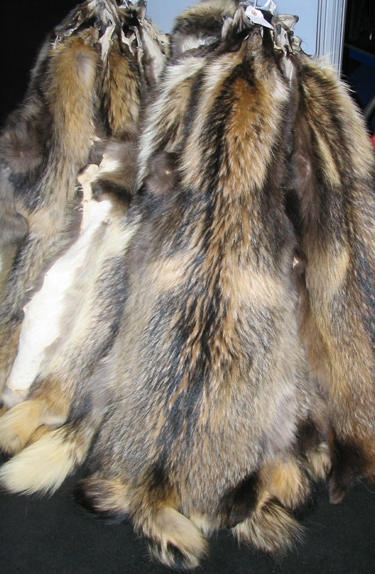
Raccoon dog pelts from the Chinese fur trade.

Being the dogs that they are, tanukis are omnivorous and will eat anything they can find, much like domestic dogs. In many areas of their range, they come into contact with humans and dwell near urban areas. This gives them access to unlimited buffets of delicious garbage, just like a real raccoon! Though they can tolerate living in close proximity to humans, raccoon dogs prefer the wilderness. They can be found in forests with dense vegetation, mountainous regions, and even along the coast.

Credit: Tambako the Jaguar

In day to day life, raccoon dogs tend to stay together and travel in pairs or small family groups. In the wild, tanukis are generally monogamous when it comes breeding time. Males and females will form a pair bond and stay together to raise their 5-7 puppies until they are ready to become independent. Raccoon dogs are altricial, meaning they are helpless when they are born. At birth, they have downy black fur and their eyes are closed. By 9 or 10 days, their eyes open and teeth can be seen on average at about 14 days. These dogs grow quickly and will reach sexual maturity by 10 months.

Like all dogs, raccoon dogs have an exceptional sense of smell. This super sense is used as a primary means of communication amongst a family or strangers. Groups of these animals will share communal “latrines” (because they are fancy) where they urinate and defecate. The scents associated with the leavings can reveal a lot about an individual dog, and this information is “read” by others like a Facebook status.
Vocal communication is also used, but, funnily enough, these dogs are the only member of their family that do not bark. Vocalizations consist of whines, mews, whimpers, and growls, which correspond to friendly, submissive, or aggressive behaviors.
So now that we’ve established how cute and awesome these creatures are, what does any of this have to do with Shigeru Miyamoto’s Super Mario Bros. franchise? I’ll tell you. The raccoon dog is actually the inspiration for the very useful Tanooki Suit that occurs in many Mario games. Although this suit is based on the Japanese mythology behind the small canid, it is often mistaken by people or mistranslated as a raccoon suit. This is not helped by the fact that the suit has a striped raccoon tail that is not shared by raccoon dogs. Japanese myths surrounding these dogs include their ability to use leaves to shape-shift and cause mischief. This is paralleled in the way that Mario can become Tanooki Mario by obtaining a Super Leaf, resulting in the ability to fly and transform into a statue to avoid or attack enemies.

Tanooki Mario.

Unfortunately, or fortunately (depends on how you look at it), real tanukis can’t fly or use leaves to transform themselves. Despite that, though, they’re pretty special pups that deserved to be acknowledged, if only for their cute little raccoon masks. From ancient legends, to Super Mario, to Animal Crossing’s Tom Nook, it’s no wonder they’ve become such an icon in Japanese culture. Animals that have such striking visual resemblance to completely unrelated species always offer something to talk about.

References:
1. Carr, Kelly and Shefferly, Nandy, ed. “Nyctereutes procyonoides”. Animal Diversity Web. University of Michigan Museum of Zoology, 2004 Web. 3 September 2015. < http://animaldiversity.org/accounts/Nyctereutes_procyonoides/>

2. Kauhala, K and Saeki, M. “Nyctereutes procyonoides (Raccoon Dog).” Red List. International Union for the Conservation of Nature, 2008. Web. 3 September 2015. <http://www.iucnredlist.org/details/14925/0&gt;.

3. “Raccoon Dog, Tanuki.” WAZA. World Association of Zoos and Aquariums, n.d. Web. 03 Sept. 2015. <http://www.waza.org/en/zoo/visit-the-zoo/dogs-and-hyenas/nyctereutes-procyonoides&gt;.

4. “Raccoon Dog – Nyctereutes Procyonoides (Eurasia’s Carnivores).” Large Herbivore Network. European Centre for Nature Conservation, n.d. Web. 3 Sept. 2015. <http://www.lhnet.org/raccoon-dog/&gt;.

5. “Tanooki Suit.” Super Mario Wiki, the Mario Encyclopedia. N.p., n.d. Web. 25 Sept. 2015. <http://www.mariowiki.com/Tanooki_Suit&gt;.

Photo Links:
1.http://www.factzoo.com/sites/all/img/mammals/raccoon-dog.jpg

2.https://flickr.com/photos/-mbender

3.https://en.wikipedia.org/wiki/File:Chinese_raccoon_fur_skins.jpg#filelinks

4.https://flickr.com/photos/tambako/

5.http://retrieverman.wordpress.com/files/2009/02/raccoon-dogs1.jpg

6.https://www.mariowiki.com/File:Tanooki_Mario_Artwork_-_Super_Mario_3D_World.png

Eight-legged, Bouncing Ball of Cute! – Peacock Jumping Spider

All Photo and Video Credits go to Jürgen Otto. Please visit his amazing website, Flickr, and YouTube accounts!
https://www.peacockspider.org/
https://www.flickr.com/photos/59431731@N05/
https://www.youtube.com/channel/UCA1shkhjIY_QiDdYfEYDqew

Peacock spiders of the genus Maratus are tiny jumping spiders barely four millimeters in length that are endemic to Australia. 53 species of Maratus spiders have been named as of 2015, 20 of which were recently discovered by researcher Jürgen Otto and his team. Though extremely small, these little jumping spiders make up for it with their spectacular colors and mating dances. It’s the males who are really the stars of the show. Females are all dull brown in color and don’t perform any sort of courtship display. They just watch and judge as the little males strut their stuff, trying to win some love.

5479964413_803612caf7_z
M. volans.

Males are equipped with elongated third legs which they wave and vibrate in accompaniment with their dance. The back of the abdomen is covered with a flap (which I like to refer to as the “butt flap”) containing brightly colored scales that form special patterns in separate species. These are held up over their heads like miniature peacock tails as they dance around in front of their potential mates. As a male dances, he slowly and cautiously approaches the female and gently places his front legs on her head. If he’s lucky, he will get to mate with her. If not, he must get away quickly to avoid being eaten. These displays can last anywhere from 20 minutes to over an hour!
Just recently, three new specie, M. jactatus, M. sceletus, and M. elephans were discovered. M. jactatus has been affectionately nicknamed “Sparklemuffin” by the graduate student who discovered it.

Sparklemuffin.

m. sceletus
M. sceletus (Skeletorus)

Peacock spiders have also been known as “flying” spiders because of their special flaps. This was because they were first falsely thought to use these flaps as wings to glide through the air, much like a flying squirrel. However, it has since been observed that they engage in no such behavior. When they jump, their flaps have no significant effects on their speed or trajectory. In fact, courtship is the only activity where these flaps are unfolded. One species, M. vespertilio, is an exception and displays his fan while sparring with other males. None of the other species have shown these male to male contests yet and this behavior was discovered accidentally while filming was taking place.

6343076673_e0a1fd2d19_z
M. vespertilio males facing off

These spiders are a perfect example of all the bizarre and amazing creature that we still haven’t found. It is amazing that something so tiny possesses such complex, fascinating, and adorable behavior. Until peacock spiders were observed, scientists had no idea that spiders displayed to each other like this. This just goes to show that there will always be more to learn and every tiny creature that we find is worth protecting.

References:
1. Girard, Madeline B., and John A. Endler. “Peacock spiders.” Current Biology 24.13 (2014): R588-R590.

2. Otto, J. C., and D. E. Hill. “Contests between male Maratus vespertilio (Simon 1901)(Araneae: Salticidae).” Peckhamia 98 (2012): 1-17.

3. Framenau, Volker W. “Australasian Arachnological Society.” (2006).

4. Arnold, Carrie. “Behold Sparklemuffin and Skeletorus, New Peacock Spiders.” National Geographic. National Geographic Society, 24 Mar. 2015. Web. 10 Aug. 2015. <http://news.nationalgeographic.com/2015/03/150324-australia-peacock-spider-sparklemuffin-new-species/&gt;.

A Local Favorite – Pacific Geoduck

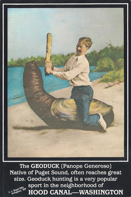
“What is a penis doing sticking out of the sand?!” you might be asking. Well, that penis is not actually a penis at all. It’s a very large and unfortunately shaped bivalve mollusk called the Pacific geoduck (Panopea generosa), or “King of Clams”. Geoducks (pronounced “gooey-duck”) are the largest burrowing clams in the world and are found exclusively in the Puget Sound region of Washington State and a southern portion of British Columbia, Canada. As baby clams, they burrow into the sediment at depths of 10-80 feet of water (but found up to 360 feet as well) and the larger they grow, the slower they burrow. Upon reaching adult size at around 3 feet long, they stay put for the rest of their lives, feeding (and squirting!) with their siphons that stick above the sand’s surface. Geoducks are among the longest lived animals, capable of living for up to 146 years!

Note: Not actual size… also, the genus name is misspelled and the species name is improperly capitalized.

Often, though, their phallic lives are cut short since they are such a coveted clam for eating. There are many geoduck farms where they are raised specifically for commercial sale, and digging for geoducks is a favorite pastime of Pacific Northwesterners. These clams are capable of withdrawing their siphons down their holes, which can be three or more feet deep, so it can be a challenge to dig for them. The animal is extricated by digging a deep hole around it with a shovel or other tool, then finishing the digging by hand and lifting the geoduck out by the shell. The legal limit for collecting geoducks in the wild is three clams per person during clam season. This might not seem like much if you think in terms of smaller bivalves like butter or razor clams, but geoducks frequently weigh a whopping three or more pounds. The record confirmed weight for this King of Clams is 8.16 pounds!

Diggin’ for Ducks

Not only is the geoduck tasty to people who like clams, it’s also the mascot of Washington’s Evergreen State College! Speedy the Geoduck was adopted by the college early on in its history and spawned the Geoduck Fight Song as well as the school motto, “Omnia Extares!”, or “Let It All Hang Out!” Speedy represents Evergreen’s most prized qualities, such as non-competitiveness, versatility and flexibility, and permanence.

IMG_2710

Speedy the Geoduck at Evergreen College’s 2014 Commencement ceremony. I took this photo when I went to my friend’s graduation.

Speedy the Geoduck was rated the “#1 worst college mascot” on many sites such as SportsPickle. The Geoducks also got rated the “#1 worst team name” from Time, and are high ranking on a multitude of other scales like weirdest mascots, most ridiculous mascots, and interesting mascots.
This is one bivalve that knows how to make a splash, whether it be in a good or a bad way, and we’re lucky to have it where I live.

References:
1. “Geoduck.” Puget Sound Shorelines. Department of Ecology, n.d. Web. 05 Aug. 2015. http://www.ecy.wa.gov/programs/sea/pugetsound/species/geoduck.html

2. “Geoduck Clams Identification & Information.” Fishing & Shellfishing. Washington Department of Fish & Wildlife, n.d. Web. 05 Aug. 2015. http://wdfw.wa.gov/fishing/shellfish/geoduck/

3. “How to Dig Geoduck Clams.” Fishing & Shellfishing. Washington Department of Fish & Wildlife, n.d. Web. 05 Aug. 2015. <http://wdfw.wa.gov/fishing/shellfish/geoduck/howto_dig.html&gt;.

4. “Geoduck.” Geoduck RSS. Geoduck.com, n.d. Web. 05 Aug. 2015.

5. Goodwin, C. Lynn, and Bruce Pease. Species Profiles. Life Histories and Environmental Requirements of Coastal Fishes and Invertebrates (Pacific Northwest). Pacific Geoduck Clam. WASHINGTON STATE DEPT OF FISHERIES BRINNON POINT WHITNEY SHELLFISH LAB, 1989.

6. “It’s Speedy the Geoduck.” Geoducks. The Evergreen State College, n.d. Web. 05 Aug. 2015. http://evergreen.edu/geoduck/home.htm

Photo Links:
1.https://commons.wikimedia.org/wiki/File:Geoduck_1.jpg

2.https://creaturefacts.org/wp-content/uploads/2015/08/f063d-geoduckpostcard.jpg

3.https://wdfw.wa.gov/species-habitats/species/panopea-generosa

The Human Centipede, Except with Worms

There are many animals that reproduce asexually – stick insects, fish, armadillos, frogs, lizards – the list could go on. When it comes to weird ways to do this, though, Aeolosoma takes the cake.
Aeolosoma is a genus of tiny, transparent worms from the class Oligochaeta, which includes earthworms and other segmented worms. They are almost exclusively aquatic and range in length from 1-10 millimeters. These oligochaetes feed on vegetation and water scum, as well as any small organisms unfortunate enough to get hoovered in with the current produced by their mouth cilia. This is all pretty typical for aquatic annelids, except when they start reproducing like a horror movie monster.

When a worm is mature, its last segment (the pygidial segment) starts to become a completely separate worm through the development of what is called a pygidial bud. Once that worm has grown all its segments, it too forms a new worm from its dividing pygidium. This continues until a chain of 2-6 worms is formed, at which point the chain divides in half and the process continues. Each of these developing worms is referred to as a zoid (plural: zooids). How does the worm behind another worm live, you ask? Well, it’s almost exactly like that disgusting movie The Human Centipede, only with worms. All of the zooids are connected through their circulatory and digestive systems and move in a coordinated way. Only when a worm is about to separate does it show independent muscle movement and only the last segment shows this.

Nightmare reproduction!

Researchers Fred Kamemoto and Clarence Goodnight  looked at the effects on Aeolosoma reproduction of different concentrations of calcium, potassium, and magnesium ions at varying temperatures. After a lot of time and a lot of worms, they concluded that there is a very complex interaction between the ions that makes it difficult for the effect of one to be singled out from any other. However, they generally found higher concentrations of calcium to be stimulating at most temperatures and higher potassium concentrations to have a toxic effect. At 10ºC, no reproduction occurred and the worms remained in a dormant state throughout the experiment. In other words, some like it hot, including these worms.
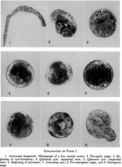
Other studies have also been done on how these little worms cope with temperature, such as Henriette Herlant-Meewis’ research on cyst formation. Cyst formation in Aeolosoma hemprichi is a process in which the worm secretes a protective mucus that hardens and encloses it in a shell-like sphere. It was first discovered in 1905 when the worms were exposed to cold, winter temperatures. Herlant-Meewis experimented with temperatures from at 3ºC, 6ºC, and 11ºC and observed their responses. Cysts did not form in worms subjected to an 11ºC environment, but many cysts were seen after 30 days in 3ºC and 6ºC medium. She took her study a step further after these observations and decided to throw food supply into the mix of cyst formation limitations.

Cyst

Cyst formation from Herlant-Meewis’ paper.

Aeolosoma were removed from their cozy, 18ºC stock culture full of nutrients and placed in pure water at 3ºC and 6ºC. By 15 days, all worms were either dead or dying and no cysts or pre-cysts stages were found. She followed up by taking 250 healthy worms living in 6ºC medium with plenty of nutrients and separated them into two groups once they had reached the pre-cystic stage. One group was placed in the same conditions as they were taken from, and the other group was subjected to pure water with no food. After seven days, Aeolosoma in the first group began to form cysts, while those in the second group died and did not form cysts.
Her conclusions were that the worms need plenty of food to store as a reserve when they enter a cyst stage. It is like a form of hibernation. If there is not a sufficient food source, they will not have the energy to form a cyst to protect themselves from the cold and will die in consequence. This is suspected to occur in the winter when water temperatures drop. Worms will feed voraciously to stock up before they form cysts and fall to the bottom of their pond or pool. Here, they can endure in warmer pockets that stay above freezing and emerge in spring as temperatures warm.

Aeolosoma worms may be small and simple creatures, but many a researcher has been smitten with their curious ways, striving to understand everything they possibly can about them. This just goes to show that even the most seemingly boring and nondescript organisms can tell us the most amazing stories about their lives if we just take the time to observe and learn.

References:
1. Kamemoto, Fred I., and Goodnight, Clarence J. “The effects of various concentrations of ions on the asexual reproduction of the oligochaete Aeolosoma hemprichi.” Transactions of the American Microscopical Society (1956): 219-228.

2. Falconi, Rosanna, Tommaso Renzulli, and Francesco Zaccanti. “Survival and reproduction in Aeolosoma viride (Annelida, Aphanoneura).” Hydrobiologia 564.1 (2006): 95-99.

3. Herlant-Meewis, Henriette. “Cyst-Formation in Aeolosoma Hemprichi (Ehr).” Biological Bulletin (1950): 173-180.

Photo Links:
1.http://www.naturamediterraneo.com/forum/topic.asp?TOPIC_ID=206115

2.http://animalkingdom.su/books/item/f00/s00/z0000048/pic/000323.jpg

3.http://www.jstor.org/stable/1538737?seq=3#page_scan_tab_contents

A Feathery Facade

Photo by Jan-Cor Jacobs

It may be hard to believe when you first look at it, but this is not a hummingbird. This is a hummingbird hawk-moth (Macroglossum stellatarum) from the Old World. It comes from the family Sphingidae, which contains the hawk moths, also called sphinx moths or hornworms. Unlike many moth species, this little one likes to be out during the day. Their hummingbird disguises are so superb that they even have tiny tails with markings on them and hold their legs against their bodies to make it appear as if they only have two. This is an excellent example of convergent evolution, in which unrelated species independently develop the same design or biological traits and fill the same niche.

Hummingbird hawk-moths feed on nectar like most other moths. Some other their favorite plants include butterfly bush, honeysuckle, foxgloves, and delphiniums. Just like hummingbirds, they hover by beating their wings with blinding speed and fly with extreme precision. The moth somehow knows not to visit the same flower twice in a row, as this would be an unnecessary expenditure of energy for nothing in return. How they keep track of which flowers they have gone to and which they have not isn’t currently known. Flying like they do takes so much energy that they must spend almost every minute feeding to replenish what they burn. Even while mating, the female moth may continue to feed.

Although it looks so bird-like in the air, the hummingbird hawk-moth looks very much like the moth that it is when perched.

A resting hummingbird moth by Christophe Quintin

The larvae, of course, are even less bird-like. It’s remarkable that this large grub turns into such a dainty and elegant little moth.

Hummingbird moth caterpillar

Hummingbird hawk-moths have relatively good vision and are able to recognize different colors quite well. Perhaps this is one of the ways they visit the individual flowers of a plant only once. They also have good navigation as they have been found to visit the same flower patches daily, similar to bees. Hummingbird hawk-moths like to fly most in bright sun, but they will also feed at dawn, dusk, and occasionally while it is raining.

Photo by Frans (fotoopa)

So next time if you are somewhere in Southern Europe, Asia, or North Africa, find some flowers and watch them to see if you can get a glimpse of this beautiful little moth.

References:
1. Jacinto, Valtar. “Macroglossum stellatarum” Encyclopedia of Life. Eol.org, n.d. Web. 25 June 2015. < http://eol.org/pages/396579/overview&gt;

2. Kelber, A. L. M. U. T. “Colour learning in the hawkmoth Macroglossum stellatarum.” The Journal of experimental biology 199.5 (1996): 1127-1131.

3. Kelber, Almut, and U. Henique. “Trichromatic colour vision in the hummingbird hawkmoth, Macroglossum stellatarum L.” Journal of Comparative Physiology A 184.5 (1999): 535-541.

Photo and Video Links:
1.https://flickr.com/photos/135003221@N02/

2.http://www.jochemnet.de/fiu/BSC1011/BSC1011_2/sld007.htm

3.https://www.youtube.com/watch?v=87EPaLYVA88

4.https://flickr.com/photos/34878947@N04/

5.https://commons.wikimedia.org/wiki/File:Macroglossum.stellatarum.caterpillar.3088.Liosi.jpg

6.https://flickr.com/photos/fotoopa_hs/

The Scaly Monogamist – Shingleback Lizard

The shingleback lizard (Tiliqua rugosa) is most at home in the rugged, dry plains and shrublands of southern and western Australia. It is a large species of blue-tongued skink that can reach a foot in length. This lizard may not look like much with its dull, armored skin and stubby tail, but it forms a bond that is quite unusual in the world of reptiles, especially among lizards.

The very head-like tail of the shingleback lizard.

Unlike almost all other lizards, the shingleback is socially monogamous. When a male and female form a pair bond, they will maintain that bond for the rest of their lives. During the breeding season, they come together and stay with each other for a couple of months and then mate. Once the female is pregnant, the two will separate, but they will reunite again the next year. These lizards have been known to remain together for over 10 years or more and may sometimes spend time together outside of breeding season as well. Shinglebacks are slow moving and often bask in the sun on or near roadways. Unfortunately, some of them get hit and killed by cars because they aren’t able to move fast enough to get out of the way. This has revealed that even after the death of one lizard, its mate continues to keep that bond. A shingleback that has lost its mate by the roadside may stay with it for several days, gently nudging it and licking it. This may be one of the closest things to grieving that has been observed in reptiles, and it is really quite touching.

51642905977_081e793f93_c

A pair of mated shinglebacks by Melissa Bruton

Though they have no defenses against cars, shinglebacks do have several ways of dealing with natural predators. One defense adaptation is their tails. Short, stumpy, and rounded, the tail looks almost exactly like the lizard’s head and may trick a predator into attacking this much less crucial part of its body, or just confuse it enough to make it leave. If this fails, they can use the threat display that is famous within the Tiliqua genus – the blue tongue. When threatened, a shingleback may raise its head, hiss, and open its mouth to reveal its blue-black tongue to scare off a predator. This startling display is followed up with a painful bite if the message isn’t clear enough. The last option for this lizard is to flatten itself to the ground and continue hissing.

8604570652_36110ea7f0_b

A shingleback showing its blue tongue by “Al and Marie” on Flickr.

Most of the time, adult shinglebacks are left alone by predators and are allowed to go about their business, such as reproducing, in peace. Shortly after mating when a female shingleback parts ways with her partner, she will produce one to four large babies. This skink is ovoviviparous, which means she gives birth to live young without connection to a placenta. The eggs stay in her body and she incubates them by collecting thermal energy from basking in the sun. When they are mature enough and have hatched, she gives birth. All the babies together can weigh up to a third of her body weight, which is the equivalent of a human mother giving birth to a three year old child. After birth, the new shinglebacks will stay with their mother for several months until they are independent enough to fend for themselves. They won’t go too far, though, as families of shinglebacks tend to stay near each other within the area.

People generally think of reptiles as cold, unfeeling, and primitive. This quiet, desert skink, and many other reptiles, prove that there is more to them than just pure, unthinking instinct. They can be gentle, sophisticated, and tender, showing many of the characteristics which we find so endearing in our warm blooded relatives. So go out and show reptiles a little love. Some of them just might be able to return it in their own, scaly way.

References:
1. Loch, Thomas. “Tiliqua rugosa (Shingleback Lizard)”. Animal Diversity Web. University of Michigan Museum of Zoology, 17 Feb. 2009 Web. 24 June 2015. < http://animaldiversity.org/accounts/Tiliqua_rugosa/&gt;

2. Bull, C. Michael, Steven JB Cooper, and Ben C. Baghurst. “Social monogamy and extra-pair fertilization in an Australian lizard, Tiliqua rugosa.” Behavioral Ecology and Sociobiology 44.1 (1998): 63-72.

3. “Shingleback Lizard.” Australian Museum. Australian Museum, 16 Dec. 2013. Web. 24 June 2015. <http://australianmuseum.net.au/shingleback-lizard&gt;.

4. Bull, C. Michael. “Monogamy in lizards.” Behavioural Processes 51.1 (2000): 7-20.

Photo and Video Links:
1.https://upload.wikimedia.org/wikipedia/commons/1/19/Shingleback_Skink_at_Sydney_Wildlife_World.jpg

2.https://flic.kr/p/2mFvoJD

3.https://flic.kr/p/2mY39fT

4.https://www.youtube.com/watch?v=F4zau3U8aGU

Little Kitty – Kodkod

If you have never loved and been loved in return by a cat or other pet, I would argue that you have never really lived. The love you feel for a cat and the love that a cat gives to you is among the truest and purest form that love can take. When you experience that love, you become a better person with a greater capacity to share that love with others in your life.
I have very recently lost the light of my life, Elly, the cat that has been with me for 18 years. She went on her own time out in the garden where she loved to be and had loving support all the way. I have never missed anyone so much and the pain of her death is still raw. But that pain comes from my love for her. I am so thankful for the time I was given with her and will always love her infinitely. She was the most special friend that anyone could ever have and I am glad that she went peacefully and didn’t have to suffer.
This is for Elly. I will love you forever, little girl!

The kodkod (Leopardus guigna) is the smallest wild cat in the Americas, and rivals several other species for the title of smallest cat in the world. This tiny cat barely reaches 6 pounds (smaller than the average house cat) and lives in the montane areas of Chile and a small portion of Argentina. It prefers dense undergrowth in its moist, temperate evergreen forest habitat, and can be found up to the tree line at around 6,300 feet. Although this cat is usually brownish yellow with black spots, melanism is common. The paws are large with black pads and its ears are rounded with a white spot on the back of each one. The kodkod’s short, bushy tail, small frame, and petite facial features give it the appearance of a kitten, even when it is a full grow adult.

There is still not much known about this small cat, and its populations are dwindling. An estimated 10,000 cats are left in the wild, no single population exceeding 2,000 individuals. Kodkods are sometimes killed for raiding chicken coops or accidentally caught in fox traps. The cats are also illegally trapped for their fur. Perhaps the biggest threat to the kodkod, though, is habitat loss and fragmentation. Kodkods don’t do well in deforested areas. They need at least a thick ground cover such as scrub to survive, and stick exclusively to the forested corridors around developed or farmed areas.

In contrast to other cats, kodkods are equally active at night and during the day. Around humans, however, they are strictly nocturnal. They appear in areas where there are high numbers of small, mouse sized prey and not many larger predators. This relative lack of competition gives them the opportunity to flourish on the abundance of food and raise their one to three kittens.

There are many conservation laws and programs in place to protect this special little cat and its close relatives like the oncilla and Geoffroy’s cat. We treat our pets as family and give them all the love we have, doing whatever we can to support them. Why can’t we show wild cats this same love? They’re really not that different.

References:
1. “Kodkod.” Felidae Conservation Fund. Felidae Conservation Fund, 2013. Web. 22 July 2015. <http%3A%2F%2Fwww.felidaefund.org%2F%3Fq%3Dspecies-kodkod>.

2. “Guigna Facts.” Big Cat Rescue. Bigcatrescue.org, 15 Mar. 2015. Web. 22 July 2015. <http%3A%2F%2Fbigcatrescue.org%2Fkodkod-guigna-facts%2F>.

3. “Kodkod.” International Society for Endangered Cats (ISEC) Canada. ISEC Canada, 2014. Web. 22 July 2015. <http://www.wildcatconservation.org/wild-cats/south-america/kodkod/&gt;.

4. Napolitano, C., Gálvez, N., Bennett, M., Acosta-Jamett, G., and Sanderson, J. “Leopardus Guigna (Chilean Cat, Guiña, Kodkod).” Red List. International Union for the Conservation of Nature, 2015. Web. 22 July 2015. <http://www.iucnredlist.org/details/15311/0&gt;.

Photo and Video Links:

1.https://flic.kr/p/2iVdFMV

2.https://www.youtube.com/watch?v=BFA6hxfnVjM

3.http://d.ibtimes.co.uk/en/full/1360845/chile-cat.jpg

It Don’t Give a Shit! – Honey Badger

Yes, it’s finally time for one of the Internet’s most popular animals – the honey badger! Made famous by this video (please note that the cobra in the video is not a king cobra).
27663138715_ebdb4d6ab7_c

The honey badger (Mellivora capensis), also called the ratel, is a large Mustelid (member of the weasel family) that lives in sub-Saharan Africa. Males can reach 11 inches at the shoulder and a little over three feet in length, while females are slightly smaller. This animal has loose skin that allows it to twist out of an attacker’s grasp easily and without damage. The skin around its neck is thicker and has evolved as a defense during fights. Not only does the honey badger have good defenses, it also has good offense. These Mustelids possess long and very powerful claws as well as a strong jaw that can even break through a tortoise’s shell. The claws, however, are used primarily for excavation of burrows for shelter. Honey badger burrows are only a few meters in length and serve as a nesting chamber. If they can, they will sometimes use abandoned aardvark or warthog burrows instead of digging one for themselves. Not much is known about honey badger reproduction, but a female honey badger’s gestation period is suspected to be approximately six months and she usually gives birth to two cubs. These cubs remain blind for a little while after they are born and are dependent on the mother for some time.

34739009921_5e50171506_c

Honey badger carrying young

Honey badgers, like wolverines and other large members of the weasel family, are very aggressive and will attack most other animals if they feel threatened. There are even incidents of honey badgers fighting against larger predators like lions or species of wild canine. Sometimes they will even steal kills from these predators. Not many other animals prey on honey badgers and they are generally left alone because they are such tireless fighters and wear opponents out easily.

When it comes to diet, honey badgers are typically carnivorous. They will eat anything they find including eggs, lizards, birds, snakes, rodents, tortoises, frogs, insects, and even sometimes dead human bodies that they dig up. Roots, berries, and other vegetation is also part of their diet, as is honey. Honey is one of their favorite foods, as the name suggests, and they will go to great lengths to destroy beehives to get to it. Though it is disputed, there are undocumented accounts of greater honeyguides (Indicator indicator) leading honey badgers to hives. Bee stings have a minimal effect this animal because its skin is so tough and impervious. This lets the honey badger eat the honey and bee larvae that it exposes at its leisure. However, there are still occurrences of badgers dying while caught in traps due the large number of bee stings they have received. It also has a reversible anal pouch that produces an extremely strong smell which is thought to calm the bees and ward off predators.

In many areas where there are large human populations, honey badgers have adapted to raid livestock and poultry. These very determined animals will rip apart chicken coops to gain access to the birds and their eggs. If caught, a honey badger will aggressively defend itself and prove very hard to kill. Not only have honey badgers become a nuisance in farming areas, but they are an excellent reservoir for the rabies virus in Kenya as well. This poses a danger to both other wild animals in the area and humans and pets who are unlucky enough to encounter a rabid badger.
Overall, the honey badger is not an animal that should be treated with nonchalance. Although it might be kind of cute, it should be given plenty of space and respect.

References:
1. Begg, Keith, and Colleen Begg. “The Honey Badger – Mellivora Capensis.” The Honey Badger. Cocopine Web Solutions, 2014. Web. 25 June 2015. <http://www.honeybadger.com/index.html&gt;.

2. Stankowich, Theodore, Tim Caro, and Matthew Cox. “Bold coloration and the evolution of aposematism in terrestrial carnivores.” Evolution 65.11 (2011): 3090-3099.

3. Kruuk, H. “Transmitting Disease to People and Livestock.” Hunter and Hunted: Relationships between Carnivores and People. Cambridge, UK: Cambridge UP, 2002. 94. Print.

4. Hamerton, Denise. “Mellivora Capensis (Honey Badger).” Biodiversity Explorer. Iziko: Museums of Cape Town, n.d. Web. 25 June 2015. <http%3A%2F%2Fwww.biodiversityexplorer.org%2Fmammals%2Fcarnivora%2Fmellivora_capensis.htm>.

5. Sheidt, Mike. “Mellivora Capensis.” Adaptations. University of Wisconsin, La Crosse, 2007. Web. 25 June 2015. <https://bioweb.uwlax.edu/bio203/s2014/scheidt_mich/adaptation.htm&gt;.

Photo and Video Links:
1.https://www.youtube.com/watch?v=4r7wHMg5Yjg

2.https://flic.kr/p/J9uPnv

3.https://flic.kr/p/UVLvmV

4.https://www.youtube.com/watch?v=iksnk1YVkac

5.https://www.youtube.com/watch?v=KSoNKpBQgJc