The Tree of Death – Manchineel

Plants can be terrifying organisms. Trees, however, are generally not often counted among those to be feared. For the vast majority of species, the only harm they are capable of inflicting is caused by falling over at an inopportune moment in time. This is wildly not the case for the manchineel tree (Hippomane mancinella L.). Native to the tropical Americas, primarily Central and South America, the West Indies, and the peninsula of Florida, this plant is commonly known as arbol de la muerte and manzanilla de la muerte, meaning the tree of death and little apple of death, respectively (Adolf & Hecker 2004; Andreu & Friedman 2012; Boucaud-Maitre et al. 2019). Within the genus name, you may notice a familiar name: hippo. This is the Greek word for horse and you might be wondering what horses have to do with a toxic plant. The second part of the genus, mane, is derived from mania, referring to madness. As a whole, Hippomane essentially means horse madness. An ancient Greek philosopher, Theophrastus, bestowed this name upon a plant native to Greece that apparently caused horses to act crazy after eating it. Later on, Linnaeus then gave this same name to the manchineel tree when he described it (Andreu & Friedman 2012). Now that we have addressed the hippo in the room, what makes this tree so damn awful?

A stand of manchineel trees. Not the red paint on some of the trunks, designating them as dangerous. (Wikimedia Commons)

Typically growing to 20 feet tall or so but capable of reaching heights as great as 50 feet, the manchineel is a member of the spurge family, Euphorbiaceae (Andreu & Friedman 2012; Hack 2026; Lauter 1952). This diverse family is famous for the latex that damaged plants produce. You may have even experienced this yourself if you have ever broken a poinsettia leaf during the holidays. It will ooze a milky sap that is slightly irritating to the skin, but nothing more harmful than that. The vast majority of the family is similar. Some, like the rubber tree (Hevea brasiliensis Muell. Arg), are even greatly beneficial to our modern lives through providing a product that has countless uses (Guerra et al. 2021; Jacob et al. 1993). Just like its cousins, the manchineel tree also produces a natural latex (Adolf & Hecker 2004; Hack 2026). But unlike other members of its family, the sap and tissues of this plant are packed with highly toxic compounds that are capable of causing damage, even death, in a wide variety of ways. The main components that make it so dangerous are Hippomanin A and a group of 18 phenolic compounds consisting of various plant toxin chemical groups (Aluja et al. 2020). Direct contact with any part of the plant can result in inflammation, burning, blistering, and formation of lesions (Blue et al. 2011; Pitts et al. 1993). If the sap gets into the eyes, it can cause severe conjunctivitis and inflammation of the cornea leading to intense pain, blurring of the vision, and potentially temporary or permanent blindness (Howard 1981; Munoz & Norton 2024; Pitts et al. 1993). I know what you’re thinking: “That’s easy! I just won’t touch it and I’ll be fine.” Unfortunately, that’s not the case. Even if you don’t directly touch the manchineel, it can still harm you. All you need to do is just be near it. There are many instances of people taking shelter from the rain under this tree, only to be severely wounded from the water mixing with the toxic sap and dripping onto the body (Blue et al. 2011). Cutting any part of the tree creates sawdust full of these dangerous compounds that can come into contact with the skin and eyes and even be inhaled. Burning the wood releases poisonous smoke that can irritate the lungs, eyes, and skin (Munoz & Norton 2024; Muscat 2019).

Leaves and flower spike (Wikimedia Commons)

The fruits of the manchineel are small, about the size of a golf ball, but they resemble a greenish-yellow apple (hence manzanilla de la muerte) and have a similar smell. They can commonly be found scattered on the ground underneath the canopy of the trees or strewn about the beaches they call home. Due to its enticing nature, many a person has mistakenly eaten the fruit. Those who have done so often report an initially pleasantly sweet taste, quickly followed by an intense, burning pain (Stickland 2000). Eating even just a small amount can progresses from pain into dysphagia, excessive salivation, nausea, vomiting, abdominal pain, and diarrhea (Boucaud-Maitre et al. 2019; Strickland 2000). It can also result in ulceration and bleeding of the mouth, esophagus, and stomach (Carroll 1952; Boucaud-Maitre et al. 2019). In some cases, and depending upon how much fruit has been ingested, symptoms can advance to changes in heart rhythm, hypotension, depression of the central nervous system, and potentially death (Hack 2026; Pitts 1993). But eating the fruit alone is not necessary to experience the horrors of the manchineel. Water saturated with leaves and branches becomes poisonous as well (Carroll 1952; Hack 2026; Pitts et al. 1993).

The apple-like fruit of a manchineel (Credit: Hans Hillewaert)

Despite its fearsome nature, the manchineel isn’t all bad. The Kalinago and other Indigenous peoples of the lesser Antilles and Caribbean have historically used the noxious sap to coat their arrows for hunting purposes (Adolf & Hecker 2004; Boucaud-Maitre et al. 2019; Munoz & Norton 2024; Muscat 2019). It is also culturally important for its use in medicines to treat a variety of ailments (Andreu & Friedman 2012; Carroll 1952; Lauter et al. 1952). The root systems of these trees also help give structure to the seashores where they occur, preventing beach erosion (Pitts 1993). Although toxic to many animals, some appear to be immune and will readily eat the fruit or use the trees as habitat (Andreu & Friedman 2012). The small green flowers of the manchineel provide nectar to native pollinators, and honeybees will even create honey from it, which is reportedly non-toxic (Adolf & Hecker 2004; Pitts 1993). Pollinators are not the only insects with a relationship to this plant. Aluja et al. (2020) have explored an extremely close interaction between the manchineel and a species of fruit fly (Anastrepha acris) that exclusively feeds on its fruit. The larvae of the fly appear to have metabolic machinery, most likely microbes, capable of breaking down the toxic compound, Hippomanin A, within the plant’s tissues. This in turn makes the fly’s larvae a suitable and safe host for the endoparasitoid wasp, Doryctobracon areolatus (Aluja et al. 2020).

A warning sign for nearby manchineel trees (Wikimedia Commons)

The manchineel’s reputation as the world’s most dangerous tree has led to its eradication across most of its native range in Florida (Andreu & Friedman 2012; Boucaud-Maitre et al. 2019; Hack 2026; Munoz & Norton 2024; Wunderlin et al. 2025). Because of this and general habitat loss within the state, it is currently listed as an endangered species and only resides in places that are still wild such as the Everglades (Andreu & Friedman 2012; Munoz & Norton 2024; Wunderlin et al. 2025). It is still locally abundant throughout much of its range outside of Florida, however, thus posing a potential threat to those who are unfamiliar with it (Botanic Gardens Conservation International (BGCI), & IUCN SSC Global Tree Specialist Group 2019). As with any organism that can be dangerous to humans, it is important not to villainize the manchineel. Yes, it is capable of inflicting massive amounts of harm; but if it is given space and treated with the respect it deserves, it poses no threat to us and provides many ecological and cultural benefits. The more we learn about it, the more secrets we may uncover as to those benefits, particularly in medicine. The manchineel is truly an incredible and fascinating plant that exemplifies the phenomenal diversity of our planet.

References:

1. Adolf, W., & Hecker, E. (2004). On the Active Principles of the Spurge Family, X. 1,2Skin Irritants, Cocarcinogens, and Cryptic Cocarcinogens from the Latex of the Manchineel Tree. Journal of Natural Products, 47(3), 482–496. https://doi.org/10.1021/np50033a015

2. Aluja, M., Pascacio-Villafán, C., Altúzar-Molina, A., Monribot-Villanueva, J., Guerrero-Analco, J. A., Enciso, E., Ortega, R., Acosta, E., & Guillén, L. (2020). Insights into the Interaction between the Monophagous Tephritid Fly Anastrepha acris and its Highly Toxic Host Hippomane mancinella (Euphorbiaceae). Journal of Chemical Ecology, 46(4), 430–441. https://doi.org/10.1007/s10886-020-01164-8

3. Andreu, M. G., & Friedman, M. H. (2012). Hippomane mancinella, Manchineel: FOR302/FR370, 7/2012. EDIS, 2012(10). https://doi.org/10.32473/edis-fr370-2012

4. Blue, L. M., Sailing, C., DeNapoles, C., Fondots, J., & Johnson, E. S. (2011). Manchineel Dermatitis in North American Students in the Caribbean. Journal of Travel Medicine, 18(6), 422–424. https://doi.org/10.1111/j.1708-8305.2011.00568.x

5. Botanic Gardens Conservation International (BGCI), & IUCN SSC Global Tree Specialist Group. (2019). IUCN Red List of Threatened Species: Hippomane mancinella. The IUCN Red List of Threatened Species. IUCN Red List of Threatened Species. https://www.iucnredlist.org/en

6. Boucaud-Maitre, D., Cachet, X., Bouzidi, C., Riffault-Valois, L., Dupuy, C., Garnier, R., & Langrand, J. (2019). Severity of manchineel fruit (Hippomane mancinella) poisoning: A retrospective case series of 97 patients from French Poison Control Centers. Toxicon, 161, 28–32. https://doi.org/10.1016/j.toxicon.2019.02.014

7. Carroll, M. N. (1952). The Pharmacology and Toxicology of Extracts, Extractives and Crystalline Substances Obtained from Hippomane Mancinella L. [Ph.D., University of Florida]. https://www.proquest.com/docview/302035970/citation/15C0955433894D24PQ/1

8. Guerra, N. B., Sant’Ana Pegorin, G., Boratto, M. H., de Barros, N. R., de Oliveira Graeff, C. F., & Herculano, R. D. (2021). Biomedical applications of natural rubber latex from the rubber tree Hevea brasiliensis. Materials Science and Engineering: C, 126, 112126. https://doi.org/10.1016/j.msec.2021.112126

9. Hack, J. B. (2026). Toxicology Answer: The Manchineel Tree. ACEP Now. https://www.acepnow.com/article/toxicology-answer-the-manchineel-tree/

10. Howard, R. A. (1981). Three Experiences with the Manchineel (Hippomane spp., Euphorbiaceae). Biotropica, 13(3), 224–227. https://doi.org/10.2307/2388129

11. Jacob, J.-L., d’Auzac, J., & Prevôt, J.-C. (1993). The composition of natural latex fromHevea brasiliensis. Clinical Reviews in Allergy. https://doi.org/10.1007/bf02914415

12. Lauter, W. M., Fox, L. E., & Ariail, W. T. (1952). Investigation of the Toxic Principles of Hippomane mancinella, L. I. Historical Review*. Journal of the American Pharmaceutical Association (Scientific Ed.). https://doi.org/10.1002/jps.3030410412

13. Munoz, M., Whitecar, S., & Norton, S. A. (2024). Beware the Manchineel: A Case of Irritant Contact Dermatitis. Cutis, 114(4), E14–E17. https://doi.org/10.12788/cutis.1123

14. Muscat, M. K. M. (2019). Manchineel Apple of Death. EJIFCC, 30(3), 346–348.

15. Pitts, J. F., Barker, N. H., Gibbons, D. C., & Jay, J. L. (1993). Manchineel keratoconjunctivitis. The British Journal of Ophthalmology, 77(5), 284–288. https://doi.org/10.1136/bjo.77.5.284

16. Strickland, N. H. (2000). Eating a manchineel “beach apple.” BMJ : British Medical Journal, 321(7258), 428. https://doi.org/10.1136/bmj.321.7258.428

17. Wunderlin, R. P., Hansen, A. R., Frank, A. R., & Essig, F. B. (2025). Hippomane mancinella / Species Page / Plant Atlas [Academic]. Institute for Systemic Botany. Atlas of Florida Plants. https://florida.plantatlas.usf.edu/plant/species/2806

Yas Queen Sting!

   Mutillidae is a fascinating family of wasps with a wide biogeographical distribution. Known colloquially as velvet ants due the fuzzy, ant-like appearance of the apterous (wingless) females (Brothers 1989; Hennessy & West 2018), these insects are primarily concentrated in tropical and subtropical regions (Lelej & Nemkov 1997). However, there is quite a large diversity in the deserts of western and southwestern North America as well (Triplehorn & Johnson 2005). Almost all of the 220 genera and over 4,600 species are solitary ectoparasitoids of ground nesting bees and wasps (Walrdren et al. 2023). This means that individual females will search out the nest of another solitary wasp or bee species and lay their eggs on or near developing young. The mutillid larvae hatch then attach themselves to and feed on the host larvae, eventually killing them. There are several species that specialize on various flies, beetles, butterflies and moths, and roaches as well (Brothers et al. 2000; Marchiori 2022).

   These diverse insects range in size from just a few millimeters, as is the case for Pseudomethoca bequaerti and Lomachaeta hicksi, to several centimeters in species like the eastern velvet ant (Dasymutilla occidentalis). There is even quite a lot of size variation within the same species, seemingly much more so than in other insects. Having processed hundreds of specimens at Washington State University, I can personally attest to this!

Lomachaeta hicksi female – note that the scale bar is only half a millimeter!

Pseudomethoca bequaerti female – only slightly larger than the L. hicksi above.

   Mutillids are also highly sexually dimorphic (Deyrup 1988), meaning the males and females look very different. Many species so much so that it is extremely difficult, if not impossible, to pair the males and females (Mickel 1974). This makes describing species and determining their relationships a real challenge for taxonomists. Females are always wingless whereas males, with few exceptions, possess wings and are fully capable of flight (Brothers & Lelej 2017).

   This dimorphism and marked difference in life history does not just make it difficult for those who study Mutillidae, it makes it difficult for the insects themselves to find mates (VanderSal-Jensen et al. 2016). As parasitoids, females roam widely in search of hosts and do not have permanent nests of their own. Males search for females primarily while in flight and it is thought that they use vision and pheromonal cues to locate potential mates (VanderSal-Jensen et al. 2016). Phoretic copulation, that in which the male mates while carrying the female as he flies, is common in Mutillidae (Marchiori 2022; Tormos et al. 2010; Waldren 2021; VanderSal-Jensen et al. 2016). Observing this behavior is one of the methods that males and females can be paired as the same species. Imagine seeing a brightly colored wasp fly by you carrying another, wingless wasp in his legs. It’s as if he’s taking her on a romantic honeymoon trip! She’ll never fully know the joys of self-powered flight, but at least she can get a taste.

   The flightless females running energetically around on the ground may be fuzzy and adorable, but possibly their most famous feature is their sting. The pain it can cause, depending upon the species, is definitely not so cute! Mutillid stings (“Sting” is the proper term for the actual structure. “Stinger” is colloquial) are the longest in comparison to their body size of any other wasp, ant, or bee group (Sadler et al. 2018). They are also very flexible and can be moved around to different angles. Only females possesses a sting because it is a structure that has been evolutionarily modified from an ovipositor, or egg-laying tube. This means that it is perfectly safe to pick up a male velvet ant. The worst he can do is pinch you a little with his mandibles and squeak at you (more on that later), both of which are pathetically cute.

A Dasymutilla male who is very upset with me holding him. You can see his mandibles open.

   The eastern velvet ant, mentioned above, is purported to be among the most painful species to get stung by, mostly due to the sheer size of the insect and the consequently large amount of venom that is delivered (Jensen et al. 2021). A popular name for the females of this species, and closely related ones such as D. klugii, is “cow-killer,” due to the myth that the sting is so painful it can kill a cow. While this is completely false, it is still no picnic to get stung by one of these fierce little ladies! Luckily, the venom is not very dangerous to us and is only meant to cause pain (Jensen et al. 2021). D. gloriosa, the beautiful and fluffy thistle-down velvet ant, has a slightly less painful sting, but still packs a punch. I can say that several of the species that live in Washington State are not at all painful, as I have personally tested this by stinging myself with them.

The sting on this Dasymutilla monticola is insanely long! Not very painful, though.

   Color is another important feature in Mutillidae and part of what makes them so distinctive from other wasps. The majority of species have aposematic coloration which warns predators of their painful sting (Lopez et al. 2021). The complexity of velvet ant coloration has led to the largest group of Müllerian mimicry rings in the world (Lopez et al. 2021), and this phenomenon has been observed in North America, Africa, and South Asia (Boutin & Vilhelmsen 2024). Müllerian mimicry is when an animal with an honest warning signal (meaning it is actually dangerous or distasteful to predators, not just pretending to be) mimics the coloration or other cue of another with an honest signal therefore amplifying the warning signal to predators in that particular region. Batesian mimicry (animals that do not possess their own defenses mimicking the warning signals or those that do) of these Müllerian mimicry complexes also occurs amongst beetles, jumping spiders, and even male velvet ants (Wilson et al. 2012). The evolution of color in Mutillidae has made them some of the most stunningly beautiful insects on the planet.

   Not only are many mutillids extremely colorful, both sexes can also stridulate. You may be familiar with cricket chirps or the clicking of a grasshopper. This is stridulation, or sound is produced by scraping two parts of the body together. Velvet ants stridulate using an organ on the abdomen to produce a squeaking sound not unlike a tiny dog toy. This sound serves as an additional warning signal to predators (Polidori et al. 2013) and a source of amusement for entomologists like myself.

   After what I have said about the stings of these insects and the bright warning colors they possess to scare predators away, I should make it clear that they are not aggressive or out to get you in any way. On the contrary, they do not want to waste their venom on you! It is quite difficult to get a female velvet ant to actually sting you and you can safely pick her up if you put your hand next to her and allow her to climb onto it. As long as you don’t squeeze her, you should likely be able to enjoy her up close and personal without a painful incident. That being said, don’t pick one up unless you accept the risk of getting stung!

Safely holding a female Dasymutilla  (most likely D. californica)!

   If you ever encounter what appears to be a weird ant covered in colorful hair frenetically scampering across the ground, hopefully now you will know what you’re looking at. It is a shame that our little sting queens, the velvet ants, often go overlooked. They are just as diverse and interesting as the more well known social Hymenopterans like ants, yellow jackets, and honey bees and they deserve some time in the spotlight.

References:
1. Brothers, D. J. 1989. Alternative life-history styles of mutillid wasps (Insecta, Hymenoptera). Alternative Life-History Styles of Animals. Springer Netherlands 35:279–291

2. Hennesy, R. D. and West, S. A. 2018. Reproductive strategies of diurnal Mutillid wasps (Hymenoptera: Mutillidae). Contributions in Science. 526:181–188.

3. Lelej, A. S. and Nemkov, P.G. 1997. Phylogeny, evolution, and classification of Mutillidae (Hymenoptera). Far Eastern Entomologist. Far East Branch of Russian Entomological Society and Laboratory of Biology and Pedology, Vladivostok 46:1-24

4. Triplehorn, C. A. and Johnson, N. F. 2005. Family Mutillidae – velvet ants. Page 548 in E. Howe, editor. Borror and DeLong’s Introduction to the Study of Insects. Seventh edition. Thomson Brooks/Cole, Belmont, California, USA.

5. Waldren, G. C., E. A. Sadler, E. A. Murray, S. Bossert, B. N. Danforth, and J. P. Pitts. 2023. Phylogenomic inference of the higher classification of velvet ants (Hymenoptera: Mutillidae). Systematic Entomology 48:463–487.

6. Brothers, D. J., G. Tschuch, and F. Burger. 2000. Associations of mutillid wasps (Hymenoptera, Mutillidae) with eusocial insects. Insectes Sociaux 47:201–211.

7. Marchiori, C. H. 2022. Associations of mutillid wasps (Hymenoptera, Mutillidae) with eusocial insects. Open Access Research Journal of Science and Technology 5:7–33.

8. Brothers, D. J., and A. S. Lelej. 2017. Phylogeny and higher classification of Mutillidae (Hymenoptera) based on morphological reanalyses. Journal of Hymenoptera Research 60:1–97.

9. Branstetter, M. G., B. N. Danforth, J. P. Pitts, B. C. Faircloth, P. S. Ward, M. L. Buffington, M. W. Gates, R. R. Kula, and S. G. Brady. 2017. Phylogenomic insights into the evolution of stinging wasps and the origins of ants and bees. Current Biology 27:1019–1025.

10. Pilgrim, E. M., C. D. Von Dohlen, and J. P. Pitts. 2008. Molecular phylogenetics of Vespoidea indicate paraphyly of the superfamily and novel relationships of its component families and subfamilies. Zoologica Scripta 37:539–560.

11. Deyrup, M. 1988. Review of adaptations of velvet ants (Hymenoptera: Mutillidae). The Great Lakes Entomologist 21:1-4.

12. Mickel, C. E. 1974. Mutillidae Miscellanea: Taxonomy and Distribution1. Annals of the Entomological Society of America 67:461–471.

13. VanderSal-Jensen, N. D., S. C. Crews, and R. G. Gillespie. 2016. Is love in the air or at first sight? Mate finding cues used by sympatric male velvet ants (Hymenoptera: Mutillidae). Invertebrate Biology 135:200–210.

14. Tormos, J., J. D. Asís, C. Polidori, A. Benéitez, and G. Storino. 2010. The Mating Behaviour of the Velvet Ant, Nemka viduata (Hymenoptera: Mutillidae). Journal of Insect Behavior 23:117–127.

15. Waldren, G. C. 2021. The velvet ants (Hymenoptera: Mutillidae): Systematics, biology, and biogeography of a little-known family. Dissertation. Utah State University, Logan, Utah, USA.

16. Sadler, E. A., J. P. Pitts, and J. S. Wilson. 2018. Stinging wasps (Hymenoptera: Aculeata), which species have the longest sting? PeerJ 6:e4743.

17. Jensen, T., A. A. Walker, S. H. Nguyen, A.-H. Jin, J. R. Deuis, I. Vetter, G. F. King, J. O. Schmidt, and S. D. Robinson. 2021. Venom chemistry underlying the painful stings of velvet ants (Hymenoptera: Mutillidae). Cellular and Molecular Life Sciences 78:5163–5177.

18. Boutin, M., and L. Vilhelmsen. 2024. Colour pattern diversity and evolution in Oriental velvet ants (Hymenoptera: Aculeata: Mutillidae). Biological Journal of the Linnean Society:blae064.

19. Wilson, J. S., K. A. Williams, M. L. Forister, C. D. von Dohlen, and J. P. Pitts. 2012. Repeated evolution in overlapping mimicry rings among North American velvet ants. Nature Communications 3:1272.

20. Polidori, C., G. Ruffato, L. Borruso, C. Settanni, and G. Pavan. 2013. Stridulatory organ and distress call in males and females of a small velvet ant (Hymenoptera: Mutillidae). Bioacoustics 22:121–135.

The Beautiful Destroyer

   The spotted lanternfly (Lycorma delicatula) is the new kid on the block and current poster child for runaway invasive pest insects in the United States. This bug, a member of the planthoppers (Auchenorrhyncha), hails from China and its surrounding countries. One of the reasons for its off the charts threat level is its polyphagous diet. The insect feeds on the phloem of plants in a broad range of taxonomic groups (Urban and Leach 2023). Many of those groups contain economically important plants, including but not limited to hops, grapes, apples, stone fruit, almonds, pine, and hardwoods like walnut (USDA 2023). The tree of heaven (Ailanthus altissima) is its preferred host. The spotted lanternfly was first detected in Pennsylvania in 2014 and it has since spread across a large portion of the eastern U.S. (New York State Integrated Pest Management Program 2023). The range is rapidly expanding and is projected to reach a majority of the country. Many quarantine zones have been established to help slow the spread (Urban 2020). Despite these efforts, the bug will almost certainly breach these boundaries and continue to grow in numbers. Although it does best with its preferred host, mentioned above, the lanternfly is capable of reproducing without access to this resource (Uyi et al. 2020). However, development time is slower and fewer egg masses are laid before the northeastern winter kills the remaining adults. But in states with warmer climates, it could potentially survive the winter and create more offspring (Uyi et al. 2020, Xin et al. 2020). This adaptability makes it an even more threatening economical and ecological pest.

The tree of heaven – a lanternfly’s favorite food (Credit: Pixbay)

  The spotted lanternfly itself is quite stunning. In adults, the forewings are leathery in texture and a pinkish grey with contrasting black spots. This color becomes criss-crossed with a mosaic of yellow veins on the distal third of the wings. Underwings are striking red and black with a white bar on each one. The first three instars of the immature stage are black with white spots and become an intense red with black and white spots by the fourth and final instar. Being beautiful does not give these insects a pass, though. Large congregations, sometimes up to 1,000 per tree (Urban 2020), in urban and suburban areas excrete large quantities of honeydew that results in sticky buildup beneath the plants on which they feed. This sugary coating encourages the growth of sooty mold that severely impacts the health of affected plants (Urban and Leach 2023). This is becoming particularly problematic in vineyards (Urban, 2020). The mouthparts of lanternflies also leave seeping wounds in plants, causing physiological stress and leaving an entry point for pathogens (Uyi et al. 2020). Controlling the dispersal of spotted lanternflies can be very difficult because of how flexible the insects are with where they lay their eggs.

Sooty mold growth caused by honeydew (Credit: Scott Nelson)

   Management strategies are evolving as more is learned about the lanternfly’s life history and ecology as it pertains to the United States. Great care must be taken when transporting goods and equipment from areas where infestations occur. Lanternflies will lay their eggs on trees, cut wood, concrete, in the wheel wells of vehicles, and on any other natural or artificial surface that is suitable (Urban and Leach 2023). Local and federal government departments have information readily available about the pestilential planthoppers and how citizens can combat them. The primary directives are to smash adults and nymphs on site, check belongings before traveling or transporting anything in and out of buildings, destroy any egg masses found, and report sightings to help track the spread (USDA 2023).

The life cycle of a spotted lanternfly (Credit: USDA)

   The standard method of agricultural and environmental industry control remains the application of chemical insecticides, particularly the neonicotinoid dinotefuran and a variety of pyrethroids. The downside to these treatment strategies is the negative effect they can have on non-target species when applied to large areas (Urban and Leach 2023). Emerging control methods that are less environmentally harsh consist of exclusion netting, targeted insecticide application, and bio pesticides such as Beauveria bassiana, a soil dwelling fungus. Other entomopathogenic fungi are currently being explored as options as well (Urban and Leach 2023). Xin et al. (2021) surveyed parasitoids of the spotted lanternfly that occur in its native range, but more research is needed to determine whether they are practical as biological control agents.

A spotted lanternfly adult with its brilliant red hind wings displayed (Credit: Chesapeake Bay Program)

   As the spotted lanternfly invasion problem continues to evolve, many challenges are arising. The use of pesticides, as mentioned, can impact unintended targets. There is also potential for the bugs to develop resistance to the chemical weapons we throw at them. Less environmentally harmful substances including neem and essential oils have shown only mild efficacy at a local level. Sticky traps encircling tree trunks sometimes kill birds and small mammals as bycatch and do not really have a significant impact on heavy populations of lanternflies anyway (Urban and Leach 2023). There is no perfect solution and management strategies will have to change along with the pest. Only time will tell what level of havoc this beautiful destroyer will wreak upon our economy, agricultural industries, and environment.

Photo Links:
1. https://jenikirbyhistory.getarchive.net/media/ailanthus-altissima-tree-of-heaven-ailanthus-nature-landscapes-a52bd0

2. https://flic.kr/p/2nChtEh

3. https://flic.kr/p/ro5GME

4. https://flic.kr/p/2eZeFLD

References:
1. Urban, Julie M., and Heather Leach. “Biology and management of the spotted lanternfly, Lycorma delicatula (Hemiptera: Fulgoridae), in the united states.” Annual Review of Entomology 68 (2023): 151-167.

2. “Spotted Lanternfly.” USDA APHIS | Spotted Lanternfly, USDA, 2023, http://www.aphis.usda.gov/aphis/resources/pests-diseases/hungry-pests/the-threat/spotted-lanternfly/spotted-lanternfly.

3. “Spotted Lanternfly Reported Distribution in the Eastern U.S.” Spotted Lanternfly Reported Distribution Map, Cornell University College of Agriculture and Life Sciences, Aug. 2022, https://cals.cornell.edu/new-york-state-integrated-pest-management/outreach-education/whats-bugging-you/spotted-lanternfly/spotted-lanternfly-reported-distribution-map. Accessed 8 Oct. 2023.

4. Urban, Julie M. “Perspective: shedding light on spotted lanternfly impacts in the USA.” Pest management science 76.1 (2020): 10-17.

5. Uyi, Osariyekemwen, et al. “Spotted lanternfly (Hemiptera: Fulgoridae) can complete development and reproduce without access to the preferred host, Ailanthus altissima.” Environmental Entomology 49.5 (2020): 1185-1190.

6. Xin, Bei, et al. “Exploratory survey of spotted lanternfly (Hemiptera: Fulgoridae) and its natural enemies in China.” Environmental Entomology 50.1 (2021): 36-45.

Don’t Say “Ugh” To THIS Slug – Nudibranchs

   Now it’s time for one of my favorite animals out there. This article has been long overdue. I’m talking about nudibranchs! The colorful, badass, and diverse group of marine slugs. But first, I have some honorable mentions. These may not be part of the Nudibranchia order, but they’re still amazing and deserve a shoutout.

   One group is Cephalaspidea (headshield slugs), which includes such animals as the cannibalistic Navanax and sand dwelling Aglaja. Next are the Saccoglossa (“sap-sucking” sea slugs), that steal the chloroplasts of the algae they eat and use them to become solar powered. New research on this group of slugs has even revealed a species, Elysia marginata, that can self-decapitate and live as an autonomous head without a heart or digestive system as it regenerates its entire body in under a month. It’s hypothesized that the slugs do this primarily to rid themselves of heavy internal parasite loads. Only younger slugs are capable of regeneration like this. Older slugs will usually die after undergoing this extreme form of autotomy (the process of voluntarily shedding a body part, usually in response to a threat). Last but definitely not least, we have the orgy loving, purple ink spewing Aplysiidae (sea hares). This family contains the largest gastropod in the world – the black sea hare (Aplysia vaccaria) – with the most massive specimen weighing just under 31 pounds and stretching over 3 feet long! What an amazing gang of critters we have here!

Internet personality and wildlife educator, Coyote Peterson, holding a black sea hare (from his YouTube thumbnail).

   Now that we’ve met some of their relatives, let’s meet the actual nudibranchs themselves. Nudis, as they are affectionately referred to by many who study and adore them, come in a couple of flavors. We have the aeolids and the dorids. We’ll start with aeolids since they are probably the most recognizable and are famous for their gorgeous, often stunningly colorful, cerata. Cerata (singular, ceras) are extensions of the animal’s back that increase surface area and aid in respiration through gas exchange. Not only this, but these structures also contain branches of the sea slug’s digestive tract. If these two things weren’t enough, the cerata also house another secret.

Cerata on the thick-horned nudibranch (Hermissenda crassicornis).

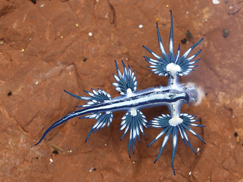
   Aeolid nudibranchs love to feed on cnidarians, a large group of animals including anemones, hydroids, corals, and jellies, and this is where their super power comes in. Famously, cnidarians sting anything they come into contact with. How do nudis get past this defensive strategy? Among other, more poorly understood modes of protection, they may use armor to keep themselves safe. Chiton, the invertebrate analogue of keratin, is notably quite tough. Some aeolids have intracellular granules of chiton both on the outside and the inside that prevent nematocysts, the packets containing coiled stinging cells, from penetrating their tissues when fired. With this ability the nudis are able to ingest the prey animal’s tentacles safely. The most impressive part about these slugs’ feeding habits is that the tip of each ceras has a structure called a cnidosac. Each cnidosac contains a few, inactivated, nematocysts that have been stolen from their prey. To summarize, aeolid nudibranchs eat their prey, stingers and all, then pass many of those stingers up to the tips of their cerata to use as their own defense. It’s very much insult to injury for the poor cnidarians they feed on. These weaponized meals are called kleptocnidae, which very fittingly means ‘stolen sting’. Some nudibranchs even possess stings strong enough to harm humans. One of these is arguably the most popular sea slug, Glaucus atlanticus (also called the blue sea dragon), probably due to its very Pokémon-like appearance. G. atlanticus is pelagic and feeds on the Portuguese man o’ war and related siphonophores, resulting in an equally potent sting.

Glaucus atlanticus photograph by Peter Woodard.

   The bright colors of many aeolid nudis are usually enough to deter predators, but if that fails, they get a nasty sting, courtesy of their own prey. Even with the warning colorations and kleptocnidae, there is the inevitability that some predators will persist. When this does occur, there are a few final parlor tricks up the slug’s proverbial sleeve. Not all nudibranchs have the ability, but many can just swim away if they’ve had enough. And remember our friend Elysia who can decapitate itself? As it turns out, plenty of aeolid nudibranchs can autotomize too, though not to the same level of drama. Many studies have demonstrated that a firm pinch to a slug’s ceras will result in its detachment, leaving the predator distracted with a squirming, mucus spewing appendage as the owner swims away. As with Elysia and many other animals that can autotomize, nudibranchs can regenerate lost cerata. It usually takes just over 40 days to fully regenerate one – a small price to pay for survival and the chance to reproduce.

An adorably plump “shaggy mouse” nudibranch (Aeolidia loui) on a shell.

   Speaking of reproduction, all nudibranchs are hermaphroditic. This means they have both male and female sex organs. Although they possess the equipment and the potential to fertilize themselves, there is little indication that they actually do so. These slugs prefer to have a partner involved. They find each other through chemicals in the trails of mucus left behind while traveling. A pair of structures on the head, the rhinophores, detect these chemicals in the water. This is akin to how a moth follows pheromones in the air with the help of its feathery antennae. The closest human analogue to this is our sense of smell, though it still differs quite radically. Many species, such as the barnacle eating dorid (Onchidoris bilamellata), often have mass spawning orgies that can cover entire rocks, but generally it is more of a quiet affair. When two slugs find each other and are both ready to mate, they participate in a circling, dance-like courtship before aligning their genitals. The reproductive opening is located on the right side of the head and contains both a penis and a vagina. One species of slug, Goniobranchus reticulatus, is famous for its disposable penis that detaches in the recipient and then regrows within a day. Plenty of others do this too, but G. reticulatus was the first to make headlines. Once mating is complete, both partners tend to go their separate ways. Sperm is stored inside a special sac until the eggs are ready to be fertilized internally. Eggs are laid in a ribbon on or near the nudibranch’s preferred prey and typically hatch within a week or 2 as tiny larvae called veligers.

Barnacle eating dorids during an orgy.

Aeolidia sp. pair with eggs.

A Heath’s dorid (Geitodoris heathi) sitting under its yellow ribbon of eggs.

   The mating behaviors discussed above are shared by both kinds of nudibranchs, but there are plenty of other things unique to the dorids. One of the most obvious of these is that they completely lack the billowing cerata that make aeolids so recognizable. Instead, they blatantly demonstrate where they get their name. Nudibranch is a mashup of Latin and Greek derived words meaning “naked” and “gills”, respectively. Cerata are multifaceted and have no single obvious function that can be visually ascertained at a quick glance and aeolids are clearly shell-less and therefore still naked as far as mollusks go. Dorids, however, display their gills right out in the open and it is very easy to recognize them as such. This tuft of gills, often referred to as “butt fluff”, is situated on top of the animal’s rear and can be drawn into the body when necessary. The gills also happen to surround the slug’s anus which… works for them. You may be wondering how these naked squishies avoid being eaten if they don’t have the fancy cerata defenses of their aeolid brethren. As it turns out, dorids have some nasty tricks of their own. Rather than stealing stingers, these not so garden variety slugs steal poisons and sequester them in their tissues. This makes them toxic. *Cue Britney Spears* A few are able to synthesize their own toxins, but most get them from prey. Dorids usually feed on sponges and can be very specialized, often only feeding on a single species. Many are very well camouflaged to their prey due to the necessity of living on or near it. Predators may have a difficult time spotting nudis like this and if they do, they may get a bad tasting surprise. Others choose to loudly advertise their toxicity with bright colors. Plenty of aeolids do this too and it is a fantastic example of aposematic coloration. “Eat me and you’ll regret it!” or “Touch me and you’ll get hurt!” These vibrant warning patterns and colors are exactly what make these animals so visually appealing to us. We see beautiful little dragons while predators see a bad time.

This thick-horned nudibranch looks like it’s glowing! You can even see its tiny eyespots.

   Nudibranchs are varied and complex animals that are continually revealing new secrets to us. If you weren’t a fan of sea slugs before, I hope you are now. It is unfortunate that their land dwelling cousins aren’t nearly as beautiful, but remember that they are still cut from the same cloth. Maybe that will change your perspective a little the next time you encounter one. Perhaps when you hear the word “slug”, your reaction will no longer be one of disgust if it was before.

Here is a link to a group of sea slug photos that show off just how diverse they are. Only one of these, the “leaf sheep” (Costasiella sp.) is not a nudibranch, although it is labeled as such: https://www.scubadiving.com/photos/photo-contest-gallery-nudibranch-central

Please also enjoy this video of slugs from the Philippines. Keep in mind, this is just one small spot, but has a mind boggling array of species. This group of animals is so insanely diverse, it’s hard not to drop your jaw in awe.

References:
1. Mitoh, Sayaka, and Yoichi Yusa. “Extreme Autotomy and Whole-Body Regeneration in Photosynthetic Sea Slugs.” Current Biology, vol. 31, no. 5, 8 Mar. 2021, doi:10.1016/j.cub.2021.01.014.

2. Rudman, Bill W. “Aplysia Vaccaria.” The Sea Slug Forum, Australian Museum, 2006, http://www.seaslugforum.net/showall/aplyvacc.

3. Greenwood, Paul G. “Acquisition and Use of Nematocysts by Cnidarian Predators.” Toxicon, vol. 54, no. 8, 6 Mar. 2009, pp. 1065–1070., doi:10.1016/j.toxicon.2009.02.029.

4. Miller, Jennifer A., and Maria Byrne. “Ceratal Autotomy and Regeneration in the Aeolid Nudibranch Phidiana Crassicornis and the Role of Predators.” Invertebrate Biology, vol. 119, no. 2, 11 May 2005, pp. 167–176., doi:10.1111/j.1744-7410.2000.tb00005.x. Accessed 22 Oct. 2022.

5. Sekizawa, Ayami, et al. “Disposable Penis and Its Replenishment in a Simultaneous Hermaphrodite.” Biology Letters, vol. 9, no. 2, 23 Apr. 2013, pp. 1–4., doi:10.1098/rsbl.2012.1150. Accessed 8 Nov. 2022.

6. Sekizawa, Ayami, et al. “Another Usage of Autotomized Penis.” Journal of Ethology, vol. 39, no. 3, 15 May 2021, pp. 319–328. Springer, doi:10.1007/s10164-021-00706-1. Accessed 8 Nov. 2022.

7. Ottuso, Patrick Thomas. “Close Encounters With the Environment.” MDedge, May 2009, cdn.mdedge.com/files/s3fs-public/Document/September-2017/083050237.pdf. Accessed 5 Nov. 2022.

8. Faulkner, D.John, et al. “Geographical Variation in Defensive Chemicals from Pacific Coast Dorid Nudibranchs and Some Related Marine Molluscs.” Comparative Biochemistry and Physiology Part C: Comparative Pharmacology, vol. 97, no. 2, 19 Apr. 1990, pp. 233–240. Science Direct, Elsevier, doi:10.1016/0742-8413(90)90133-t. Accessed 5 Nov. 2022.

Photo Sources:
1. https://www.youtube.com/watch?v=15I8eIqh9iI
2.
https://commons.wikimedia.org/wiki/File:Glaucus_atlanticus_Long_reef.jpg
All other photos  taken by me.

Video Link: https://www.youtube.com/watch?v=_f3GpKiaQUA

Ants in Your Pants

   Ants. Can’t live with them, can’t live without them. They’ve been around for ages and outnumber us by over a million to one. They’re not just home invaders and picnic crashers; they’re the only other animal known to have sophisticated agriculture and manage herds of livestock. With more than ten thousand species to choose from, there’s an ant for every occasion. Did you rip your favorite shirt? Make friends with some weaver ants. Are you looking for someone to mow your lawn? Hire an army of grass cutter ants. Is life dull and you just want a thrill? Try bothering a stampede of African driver ants and get the adrenaline rush of a lifetime. I’ve mentioned ants before, but now is their time to have the spotlight.

27714515471_49022e674f_k

African driver ants, or Siafu, are infamous for their nomadic colonies that devour anything and everything in their path. Blind soldiers with massive jaws guard the perimeters as the workers march on, carrying the young and the queen. Photo by Peter Steward on Flickr.

   I couldn’t possibly cover every ant I would like to in this article, but I did manage to choose a select few, starting with the infamous bullet ant (Paraponera clavata). This ant is well known to have the most painful known insect sting in the world. As its name infers, the sting is likened to having a bullet wound. Justin Schmidt, a dedicated and somewhat eccentric entomologist, has painstakingly (pun intended) stung himself with as many insects as possible to compile the Schmidt Sting Pain Index. The pain of various insect stings, which he describes in wildly imaginative detail, are ranked from 1 to 4 (with 4 being the most painful). Can you guess who won that coveted number 4 on the scale? Take a moment. I’ll wait. Okay, now that you’ve guessed I can confidently assume that you thought about the bullet ant. After all, it is the star of this section. If you did choose the bullet ant, you’re wrong. That honor actually goes to the tarantula hawk wasp. No, the bullet ant doesn’t play by the rules. It has a sting so awful that it got its very own ranking of 4 plus. Not even the scale could contain the pain. As with many other animal toxins, bullet ant stings affect voltage gated sodium channels on cell membranes. This can wreak havoc on a cell’s ability to transmit signals, especially in the central nervous system (CNS). The blocked signals in nociceptors and skeletal muscles are what leads to the intense pain, paralysis, uncontrollable shaking, nausea, and more. The legendary sting of P. clavata is the reason it has become the gatekeeper of boys on their way to becoming warriors. The Sateré-Mawé tribe of Brazil has used this ant for years as part of a culturally important initiation rite. One sting is more than enough to cope with, but Mawé boys must endure multiple stings. Bullet ants are collected and dipped into a natural sedative to be safely woven into large gloves made from palm leaves. At the start of the initiation, a young man must slip both his hands into gloves filled with, now irate, immobilized ants. Only a thin layer of charcoal protects his hands and he must wear these gloves for five minutes straight and take the pain. What’s worse is the effects of the venom last well beyond these five excruciating minutes and boys hoping to become warriors must undergo this nightmare a total of twenty times before they are fully initiated. Forget jaguars, caimans, and anacondas. The Mawé people consider this humble ant to be the worst their rainforest home has to offer.

Paraponera clavata

Great bullet ant photo by Graham Wise on Flickr.

    Despite the potency of the venom delivered by the ant itself, one component may have medical significance. This compound of P. clavata, known as poneratoxin (PoTX), can have an analgesic effect in rats if administered in a small, controlled amount. Not only that, but it could potentially serve as a biological insecticide thanks to its ability to block transmission in the insect CNS.

   Now it’s time for an ant that poses no threat to people, but is an amazing survivor. Meet the Saharan silver ant (Cataglyphis bombycina). This ant is capable of surviving conditions that would cook virtually any other living creature. In the Sahara desert, the surface of the sand can reach temperatures of up to 158℉ under the brutal midday sun. Several ingenious evolutionary adaptations give C. bombycina the ability to cope with this unforgiving environment while all others must either hide or perish. One strategy is to limit the time of exposure. Even these hardy ants will die if they are out on the scorching sands for long enough. That’s why they turn their foraging trips into a mad dash for their lives. Groups of ants leave their nest in a frantic search for other arthropods, such as desert beetles, that were too slow to escape the sun’s deadly rays. Once located, prey is dragged back to the nest as fast as possible via extraordinary teamwork. The poor animals that die and end up providing for these ants have succumbed to what is known as heat shock. Every living organism produces substances called heat shock proteins (HSPs). HSPs are tied to specific genes (heat shock genes) that, when activated by high temperature, are expressed and direct synthesis of HSPs by the cells. The purpose of these proteins is to protect the body from severe heat shock. Normally they are not produced until there is an environmental trigger, but Saharan silver ants start to produce them before leaving their nests. HSPs are synthesized preemptively to give the ants protection so they don’t die upon exiting their nest. This is because their foraging trips are usually too short to trigger synthesis of HSPs the normal way and the temperature rise would kill them before they got the chance. Not only are these HSPs produced before exposure, they continue to be synthesized beyond temperatures that would kill other animals, giving silver ants an even greater edge.

   Another advantage over everyone else is long legs and a fast gate. Even though the sand itself may reach temperatures past what C. bobmbycina can tolerate, it is significantly cooler just a few millimeters off the surface. The long, stilt-like legs of this ant raise it off the sand just enough to keep its body below its critical maximum of 128.5℉. Long legs generally mean an animal can run faster, and C. bombycina is no exception. Its gait is a frenzied sprint that, at top speed, becomes quadrupedal. That’s right, these ants will actually lift their front legs off the ground completely to pelt forward as quickly as they can, and to minimize exposure to the sand. Running fast and limiting each foot’s contact with the ground allows them to go farther and keep cooler in their short bursts of foraging. They use the sun to navigate so that they can keep track of where they are in relation to their nest and how long they have been out.

   Unfortunately, special proteins, great legs, and excellent navigational ability aren’t enough to beat the heat. Silver ants actually spend the majority of their scavenging forays offloading heat while sitting atop dry vegetation or stones. What allows them to do this so efficiently is the hair that gives them their name. These gleaming, silvery coats are what put them above and beyond everyone else when it comes to desert survival. Each individual hair is a triangular shape with two ridged surfaces and one smooth surface. The smooth surface sits against the body and the ridged surfaces face outward. When solar radiation hits the surfaces of these hairs, the ridges trap it and scatter that radiation back out in all directions. This is called “Mie scattering” and enhances the hairs’ reflectivity in the visible and near infrared range where solar radiation is at its strongest. In the mid infrared, the silvery hairs become antireflective and help the ants offload heat through emissivity. But what about the heat radiating off the ground? C. bombycina is prepared for that too. The undersides of their bodies are bare and reflect the mid infrared radiation from the sand better than if they were covered in hairs. All of these features, and more, combine to optimize survival and allow silver ants to be the lean, mean, foraging machines that they are.

   There are countless ways to protect yourself from all kinds of threats if you’re an ant. Wearing what is essentially a heat shield or having a sting that will cripple your attacker with pain are just two. How about being heavily armored and using your head as a door to seal everyone out of your house? Using your own body as a barrier is called phragmosis and it’s what several genera of ants choose to do. Ants, as colonial animals, all have specific jobs to help their family. For ants in these phragmotic genera, usually the soldiers, that job is protecting the nest with their heads when the colony is threatened. The head structures, or cranial discs, that aid this can come in a variety of shapes and sizes that match their purpose. Some are even covered with sticky, fibrous secretions that accumulate environmental debris to help escape detection (cryptic phragmosis). Not only can they be used for blocking entrances, they can also be used like bulldozers to physically push intruders out of the nest. In one species of ant, Blepharidatta conops, it is the queen who possesses a head disc that she uses to block her nest chamber. The diameter is much too small to be of use blocking the nest entrance, but it certainly gets the job done when it comes to protecting her young. Perhaps one of the most diverse phragmotic genera is Cephalotes, also called turtle ants. In Cephalotes, generally all castes from workers to queens possess some kind of highly adapted head shield. These arboreal ants dwell high in the canopies of neotropical forests and use their heads to plug the holes of their wood cavity nests. They are also capable of gliding down from the canopy using what is known as Directed Aerial Descent, which is pretty cool. These ants are basically weird insect superheroes. Anyway, the morphology of the cranial discs prevents exposure of eyes, antennae, and mandibles to predators, just leaving them with a perfectly fitted, armored door to deal with. As mentioned before, these discs can be covered in organic material that sticks to glandular secretions, making entrances hard to locate in the first place.

USA, TX, Gonzales Co.24-v-2016

For some really breathtaking photography of Cephalotes, please check out Alex Wild.

   In a fantastic example of convergent evolution, there is a genus of Old World ant, Carebara, that has independently developed a very similar head shape to that seen in Cephalotes. The convex, shield-like head disc shape may have evolved independently even among separate species of Carebara. As ground nesting ants found in the leaf litter of African forests, Carebara workers have cephalic discs that collect soil and other debris, thereby camouflaging the nest entrances. They do not possess the sticky secretions of Cephalotes, but litter and soil easily collect on small hairs. When threatened by an intruder, they lock their heads down, making them both harder to see and the nest more difficult to invade.

400px-Fischer_et_al_2015_F1

Figure 1. from Fischer et al. 2015. The phragmotic workers of two Carebara species.

   Reverse phragmosis has also been hypothesized, but as of now not proven, in the queens of the species Pheidole embolopyx. This is when the ant has a truncated (shortened), shield-like abdomen. So basically it’s a butt door. Or it would be if it was used to block nest tunnels. In the numerous experiments performed by E.O. Wilson and B. Hölldobler, not once did the study queen use her abdomen defensively. She always went on the attack, using her mandibles to nip at any threat they presented her with. She even turned completely around in her tunnel to attack the dissecting needle approaching her from behind rather than using her abdomen as a shield. It’s still a mystery what purpose this flat and potentially phragmotic abdomen serves. However, it does really reinforce the saying “Don’t judge a book by its cover.”

   That goes for any of the ants in this article or anywhere in the world. Ants have a bad reputation and we’re all too quick to judge them and make assumptions based on the bad experiences we’ve had with a handful of species. In reality, there are trillions more ants out there that are just going about their own business, often in a truly fascinating manner, and want nothing to do with us. Let’s appreciate and think about those ants before we go judging any ant we see.

The wonderful Justin O. Schmidt sadly left us in February of 2023. He was an invaluable and pioneering contributor to the scientific community and will be dearly missed.

The “King of Sting”

References:

1. Rykaczewska-Czerwinska, Monika, et al. “Antinociceptive Effect of Poneratoxin (PoTX) in Rats.” Pestycydy, no. 1-2, 2008, pp. 135–141.

2. “Sateré-Mawé Initiation.” Culture and Customs, Penn State and WordPress, 16 Nov. 2015, 1:15pm, sites.psu.edu/mgeitnerrcl/2013/11/16/satere-mawe-initiation/.

3. Torres, A.f.c., et al. “Molecular Pharmacology and Toxinology of Venom from Ants.” An Integrated View of the Molecular Recognition and Toxinology – From Analytical Procedures to Biomedical Applications, 2013, doi:10.5772/53539

4. Palma, Mario Sergio. “Insect Venom Peptides.” Handbook of Biologically Active Peptides, 2006, pp. 389–396., doi:10.1016/b978-012369442-3/50059-3.

5. el-Showk, Sedeer. “Agriculture in Ants and Humans.” Scitable, Nature Publishing Group, 17 June 2013, http://www.nature.com/scitable/blog/accumulating-glitches/ant_agriculture.

6. Moore, Hannah. “Are All the Ants as Heavy as All the Humans?” BBC News, BBC, 22 Sept. 2014, http://www.bbc.com/news/magazine-29281253.

7. Gough, Zoe. “The World’s Most Painful Insect Sting.” BBC Earth, BBC, 13 Mar. 2015, www.bbc.com/earth/story/20150312-the-worlds-most-painful-insect-sting.

8. Gehring, Walter J., and Rüdiger Wehner. “Heat Shock Protein Synthesis and Thermotolerance in Cataglyphis, an Ant from the Sahara Desert.” Proceedings of the National Academy of Sciences, vol. 92, no. 7, 28 Mar. 1995, pp. 2994–2998., doi:10.1073/pnas.92.7.2994.

9. Zollikofer, Christoph P. E. “Stepping Patterns in Ants II. Influence of Body Morphology.” Journal of Experimental Biology, vol. 192, no. 1, 15 July 1994, pp. 107–118.

10. Shi, Norman N., et al. “Keeping Cool: Enhanced Optical Reflection and Radiative Heat Dissipation in Saharan Silver Ants.” Science, vol. 349, no. 6245, 18 June 2015, pp. 298–301., doi:10.1126/science.aab3564.

11. Alonso, José Ramón. “Flat Heads.” Mapping Ignorance, Dinahosting, 8 Jan. 2016, mappingignorance.org/2016/01/08/flat-heads/.

12. Brandão, C. R. F., et al. “The First Case of Intranidal Phragmosis in Ants. The Ergatoid Queen of Blepharidatta Conops (Formicidae, Myrmicinae) Blocks the Entrance of the Brood Chamber.” Insectes Sociaux, vol. 48, no. 3, Sept. 2001, pp. 251–258., doi:10.1007/pl00001774.

13. Fischer, Georg, et al. “Two New Phragmotic Ant Species from Africa: Morphology and next-Generation Sequencing Solve a Caste Association Problem in the Genus Carebara Westwood.” ZooKeys, vol. 525, 2015, pp. 77–105., doi:10.3897/zookeys.525.6057.

14. Yanoviak, Stephen P., et al. “Evolution and Ecology of Directed Aerial Descent in Arboreal Ants.” Integrative and Comparative Biology, vol. 51, no. 6, 2011, pp. 944–956., doi:10.1093/icb/icr006.

15. Wilson, E. O., and B. Hölldobler. “Caste-Specific Techniques of Defense in the Polymorphic AntPheidole Embolopyx (Hymenoptera: Formicidae).” Insectes Sociaux, vol. 32, no. 1, 1985, pp. 3–22., doi:10.1007/bf02233223.

Photo and Video Links:

1. Guarding the empire’s treasure by Peter Steward

2. Paraponera clavata by Graham Wise

3. https://www.youtube.com/watch?v=VbkgNDERlpU

4. Texas Turtle Ant, solider (Formicidae, Cephalotes texanus) by Alejandro Santillana

5. Figure 1

6. https://www.chemistryworld.com/careers/the-man-who-gets-stung-by-insects/2500173.article

Otterly Adorable

Otters! Who doesn’t love ‘em? From the fluffy and adorable sea otter to the giant Amazonian river otter; there’s an otter for every occasion! Most are fairly social, but the Asian small-clawed otter (Aonyx cinerea) beats all the others out when it comes to gregariousness. It is the most social of all the world’s 13 otter species, forming lifelong pair bonds that lead to extended family groups of as many as 15 or even 20 individuals. In addition to being a family oriented otter, it’s also the chattiest; you have to be to communicate with your many relatives. Along with the classic mustelid trait of scent marking, sound is one of the primary ways in which this animal discusses the intricacies of life. Small-clawed otters are known to use a repertoire of at least 12 different kinds of vocalization including chirps, squeals, barks, and shrieks. They’re like a group of excited tweens who won’t shut up about whatever boy band is popular with the kids these days. Unlike tween girls, though, the small-clawed otters’ company is actually enjoyable because they’re so darn cute and fuzzy!

9174391704_f09ef6ce44_z

An adorable Asian small-clawed otter by David Ellis on Flickr.

Native to southern and eastern Asia, these little rascals thrive near clean freshwater streams and rivers with lots of vegetation. Occasionally, they can be found in rice paddies or along the coast near saltwater. Although small-clawed otters need a water source, they are happy to spend time on land as well. Their paws are only partially webbed due to this more terrestrial lifestyle and they use them to search for and capture prey both in the water and on land. Just like raccoons, small-clawed otters hold whatever they are eating in their delightful little hands. Sometimes they can be a nuisance in rice paddies because they will pull up rice plants while on the hunt. As mustelids, otters are playful and mischievous by nature after all.

14414655148_eb67ac283a_z

Another fantastic small-clawed otter photo by David Ellis.

Unfortunately, their numbers are currently on the decline because of habitat destruction caused by land development and competition with humans for food resources. The favorite foods of these otters, which include crabs, mussels, and clams, are also harvested by people and can be overexploited. In addition to reduction of food biomass, there is more frequent chemical contamination of these food sources from human activity. Since small-clawed otters are considered to be a vulnerable species, they are protected throughout much of their range. This is a good start, but areas that were once home to these otters have still seen them disappear.

One such case comes from Singapore. Small-clawed otters were historically common there, but are now only visitors from its offshore islands rather than permanent residents. Fortunately, though, Singapore has not remained an otter free zone. Thanks to pollution cleanup efforts in local waterways, smooth-coated otters (Lutrogale perspicillata) have recently made a return. These much larger cousins of the small-clawed otter now number over 60 individuals and can often be seen and photographed by locals and tourists alike… or so it was thought! Recent research indicates that these water weasels are not actually smooth-coated otters. As it turns out, they are a hybrid between smooth-coated and small-clawed otters. This is the first recorded observation of a wild otter hybridization.

31091942_1959808447425027_8993325990683869184_n

A family of Singaporean otters from OtterWatch’s Facebook. Go check them out!

The story behind these fuzzy hybrids is even more interesting. The research team, led by Beatrice Moretti, came up with several reasons why this union occurred and settled on the “sexual selection hypothesis” as the most likely. Years ago when small-clawed and smooth-coated otter populations overlapped in Singapore’s waters, there may have been a dearth of small-clawed males. The smooth-coated otter was more common in this region, so small-clawed females likely would have interacted with them. Initially, these females may have rejected the smooth-coated males while they searched for males of their own species. Upon being unsuccessful, the females might have settled for the smooth-coated males instead. So why were the frisky locals seen today mistaken for smooth-coated otters in the first place? Well, it’s because they look exactly like smooth-coated otters. The initial objective of the study mentioned above was to learn about smooth-coated otter populations around the world, not phylogeny and hybridization. So naturally, the researchers were confused. They thought the samples collected from their smooth-coated otters might have been contaminated by small-clawed otters swimming by for a visit! After rerunning tests and making sure that no mistakes had been made, they realized that what they found was genuine and set out to figure out why and how. This hybridization happened so many generations ago that the offspring of the first few couples who bred ended up back-crossing with smooth-coated otters. The otters that now live in Singapore contain small-clawed DNA, but superficially appear to be smooth-coated otters because of the continued back-crossing with that latter species.

18791236193_f10ce2f8a4_z

A smooth-coated otter by Marie Hale on Flickr.

Examples like this prove that even when we think we know something, nature always has surprises for us hidden up its sleeves. Perhaps with continued conservation efforts and habitat restoration, small-clawed otters will return to the waters of Singapore to frolic with their not so distant cousins.

References:
1.Hamman, David. “Aonyx Cinerea.” ADW, Animal Diversity Web, 2004, animaldiversity.org/site/accounts/information/Aonyx_cinerea.html.

2.Wright, L., et al. “Aonyx Cinereus” IUCNRedlist, The IUCN Red List of Threatened Species, 2015, http://www.iucnredlist.org/details/44166/0. Wright, L., de Silva, P., Chan, B. & Reza Lubis, I. 2015. Aonyx cinereus. The IUCN Red List of Threatened Species 2015

3.Scheifele, Peter M., et al. “Vocal Classification of Vocalizations of a Pair of Asian Small-Clawed Otters to Determine Stress.” The Journal of the Acoustical Society of America, vol. 138, no. 1, 2015, doi:10.1121/1.4922768.

4.Hong, Jose. “Surprising Branch in Singapore’s Otter Family Tree.” The Straits Times, 14 Jan. 2018, www.straitstimes.com/singapore/surprising-branch-in-otter-family-tree?xtor=CS1-10.

5.Moretti, Beatrice, et al. “Phylogeography of the Smooth-Coated Otter (Lutrogale Perspicillata): Distinct Evolutionary Lineages and Hybridization with the Asian Small-Clawed Otter (Aonyx Cinereus).” Scientific Reports, vol. 7, 27 Jan. 2017, doi:10.1038/srep41611.

Photo Links:
1.https://flic.kr/p/eYHa2j

2.https://flic.kr/p/nXLSM3

3.https://www.facebook.com/OtterWatch/?ref=page_internal

4.https://flic.kr/p/uCvZQV

Curious Cuttles – Dwarf Cuttlefish (and friends)

    There appears to be some unspoken rule that tiny versions of things are exponentially more adorable. Take for instance hummingbirds, pygmy marmosets, dwarf elephants, pygmy hippos, or Brookesia micra (the chameleon that fits comfortably on the head of a match). The dwarf cuttlefish (Sepia bandensis), with a mantle length of under three inches, is no exception. Dwarf cuttlefish are at home in the warm waters of the Indo-Pacific including, but not limited to, the Philippines, New Guinea, and Sulawesi. Living mostly in shallow coastal waters, these tiny cephalopods are most active at night, feeding on small crustaceans or fish found over sand and reef. Like almost all members of their group, dwarf cuttlefish are exclusively shallow water inhabitants. This is because of the vestigial cuttlebone that’s contained within the mantle. The cuttlebone is a remnant from the ancient history of cephalopods. Like the ancestral shell, it still retains tiny chambers filled with gas that assist with buoyancy. As a consequence, the cuttlebone will implode if the poor creature swims too deep. Being relatively restricted to shallow water does have its perks, though. Since there is so much light, cuttlefish get to fully utilize their incredible skin to create a wide variety of colors, patterns, and textures to communicate with each other and other animals.

     One of the most spectacular displays used by these tiny cuttles is known as the “passing cloud”. It consists of dark bands of color moving down the animal’s mantle via the pulsing of chromatophores in the skin. While this occurs, the cuttlefish keeps the rest of its body’s color and pattern static (unchanging).

Here you can see the passing cloud display in action on this dwarf cuttlefish that I filmed at the Seattle Aquarium. It does it at the beginning and the end of the video.

S. bandensis is not the only cuttlefish species to display these strange, psychedelic waves. Many other cuttlefish do this as well, and also some octopuses. Not surprisingly, the display differs in each species and there are many variations of it. These bands can occur on almost any part of the body and go in different directions depending on the species. The delightfully named Wunderpus photogenicus, an octopus, pulses dark bands over its eyestalks. Perhaps the most interesting thing about pulsing displays like the passing cloud is that no one knows for sure what the purpose is.

Did you notice the pulses on Wunderpus’s eyestalks?

     There is a myriad of possibilities of what the displays could be used for. Some cephalopods, such as the broadclub cuttlefish (Sepia latimanus), almost certainly use these displays for hunting. These cuttlefish can sometimes be seen rapidly pulsing the chromatophores on their arms before pouncing on prey. It’s possible that the chromatic pulses mesmerize prey, holding them in place while the cuttlefish positions itself for a deadly strike. The Australian giant cuttlefish (Sepia apama) has been observed using pulse displays in spawning aggression and while drifting in and out of seaweed. These are both notable examples of how pulse displays can be used. Male cuttlefish are well known for displaying aggression to rivals with one half of the body, while showing receptivity to females with the other half. Communication is extremely important among cephalopods and pulse displays seem to serve this purpose well. Camouflage is also a famous attribute of cuttlefish and octopuses alike. Creating rhythmic waves that mimic the rippling light coming from the surface while drifting with the weeds is an excellent way to hide. It definitely puts those chromatophores to good use by helping protect the animal from predators.

This broadclub cuttlefish is using its chromatic pulses to hypnotize a crab. It has its two outer arms positioned in what is called a “branched coral” pose.

     The functions of chromatic pulse displays like the passing cloud are not as obvious in other contexts. The extraordinarily cute and adorably named flamboyant cuttlefish (Metasepia pfefferi) will pass waves over its body in as simple a situation as sauntering over an open mudflat. There is a possibility that, along with its bright colors, this could serve as a warning to potential predators. Cephalopod expert Mark Norman has found that the flesh of this little cuttlefish is incredibly toxic. However, this is only anecdotally recorded in the NOVA special Kings of Camouflage and there has been no scientific study published yet that analyzes the toxins. Toxic or not, the flamboyant cuttlefish uses it’s chromatophores to send a message, supporting the potential communication function of pulse displays.

This tiny flamboyant cuttlefish is too cute as it walks around strutting its stuff. Could it be saying “Don’t eat me, I’m toxic!”?

     It’s pretty clear that passing clouds and other pulses have specific uses depending upon the environmental or behavioral context of the animal creating them. That is, it’s pretty clear for most of the cuttlefish I’ve mentioned. Alas, the star of this article – the diminutive dwarf cuttlefish – still hides its secrets. Although I am privileged to have seen and video recorded the passing cloud behavior at the Seattle Aquarium many times, I still can’t figure out what these little cuttles are using it for. One minute, an individual may be hovering in place with wave after wave passing over its mantle and the next it will be almost completely black and fighting with a tank mate. From my pathetically inferior human perspective, it seems random. There is the possibility that these captive bred cuttlefish are behaving a little differently than they would in the wild, or that they are influenced by the presence of people. With so many variables, it’s difficult to tell what the dwarf cuttlefish are using it for without a full blown scientific study. Even with the most skilled researchers giving it their all, we will probably never know just what goes on in the minds of these tiny cuttlefish (or any of the other species).

fullsizeoutput_c68

This is a photo I took at the Seattle Aquarium. You can see the tiny suckers on this dwarf cuttlefish’s adorable little arms as it watches me through the glass.

     Cephalopod nervous systems are so different from our own that we can only make feeble guesses at how they think and feel about the world. Can you imagine having your brain directly connected to your skin so you could change color and texture in fractions of a second just by thinking? Me neither, but wouldn’t it be cool? That’s an everyday reality for almost all modern cephalopods and something we can’t even begin to relate to. As our understanding of these fascinating creatures improves and science gives us new ways of studying them, we may come closer to discovering what it all means. Until then, let’s just admire their beautiful and complex alien language for what it is – one of the many wonderful mysteries of the natural world.

References:
1. Mustain, Andrea. “World’s Tiniest Chameleon Discovered.” LiveScience. Purch, 14 Feb. 2012. Web. 12 July 2017

2.“Stumpy-spined Cuttlefishes, Sepia bandensis.” MarineBio.org. MarineBio Conservation Society, n.d. Web. 2 March 2017.

3. How, Martin J., et al. “Dynamic Skin Patterns in Cephalopods.” Frontiers in Physiology, vol. 8, 2017, Accessed 31 July 2017.

4. Cuthill, Innes C. “Animal Behaviour: Strategic Signalling by Cephalopods.” Current Biology, vol. 17, no. 24, 2007, pp. 1059–1060. ScienceDirect, Accessed 31 July 2017.

5. Hanlon, Roger. “Cephalopod Dynamic Camouflage.” Current Biology, vol. 17, no. 11, 2007, pp. 400–404. ScienceDirect, Accessed 31 July 2017.

6. Osorio, Daniel. “Cephalopod Behavior: Skin Flicks.” Current Biology, vol. 24, no. 15, 2014, pp. 684–685. ScienceDirect, Accessed 31 July 2017.

7. Laan, Andres, et al. “Behavioral Analysis of Cuttlefish Traveling Waves and Its Implications for Neural Control.” Current Biology, vol. 24, no. 15, 2014, pp. 1737–1742. ScienceDirect, Accessed 31 July 2017.

8. Kaufmann, Gisela, director. Kings of Camouflage. NOVA, 2011.

9. Staaf, Danna. “Sheathing the Shell.” Squid Empire: the Rise and Fall of the Cephalopods, ForeEdge, an Imprint of University Press of New England, 2017, p. 112.

Video Links:
1.https://www.youtube.com/watch?v=u76P3gCLWn4

2.https://www.youtube.com/watch?v=baNp8bgOtok

3.https://www.youtube.com/watch?v=qjloPutUCeo

4.https://www.youtube.com/watch?v=51jB8YljliM

Unfishy Fish

     What do you think of when someone says “fish”? The image of a salmon or goldfish probably pops into your mind. Or perhaps a colorful tropical reef fish. Odds are, you’re not thinking of a lipstick wearing, pancake shaped animal with frog feet and a long, pointy nose. You’re most likely not thinking of a Toblerone shaped shark with swirly nostrils either. The fact is, evolution doesn’t care what your idea of a fish is. It’s going to go with what’s successful and if it looks like a nightmare clown, so be it. Evolution doesn’t have a goal or a grand plan. What happens will happen and if something works, it persists.

fishdiagrams1

A generalized fish body plan.

     So here we are with and ocean of oddities to explore. To start with, let me introduce you to something that looks more or less like a ‘proper’ fish, but didn’t get the memo about how to swim like one. Enter, the razorfish (Aeoliscus strigatus). This fish is elongated and laterally compressed, giving it a razor thin appearance, hence the name. It feeds on tiny zooplankton such as brine shrimp as it hides among corals and seagrasses. Razorfish swim vertically in small schools with their snouts pointed down at all times. It is not known exactly why they do this, but it’s certainly entertaining to watch. With razorfish, it’s always a synchronized swim show. For the razorfish, the warm waters of the western Indo-Pacific are the perfect place to engage in these underwater ballets.

poisson-rasoir-01

A school of razorfish.

     Another weird, warm water inhabitant to be featured here is at home in the lower estuary of Derwent River in Tasmania and several other locations along the southeastern coast of Australia. Unfortunately, it is now critically endangered because of a multitude of factors including a low reproduction rate, habitat destruction, and suspected predation by an introduced sea star. The spotted handfish (Brachyonichthys hirsutus) has a sail like dorsal fin and large, webbed ‘hands’ which it uses to walk along the silty bottom. It belongs to of the order Lophiiformes, along with other strange members like deep sea anglerfishes and frogfish.

spotted-handfish

Spotted handfish.

Unlike many other fish, which hatch out as larvae that further develops outside the egg, spotted handfish emerge as tiny, fully formed versions of their parents. Rather than floating among the plankton of the open water, these babies stick to the sandy floor and live out the rest of their lives there. The peculiar reproduction habits of this fish are exactly what make it vulnerable to the northern Pacific sea star (Asterias amurensis). Before they become fish, the eggs are attached to stalked sea squirts and other vertical organisms that are extremely appetizing to the star. As a consequence, the eggs are devoured along with the sea star’s target prey. Conservation efforts have since been set in motion to save this unique Australian animal. Along with a captive breeding program and hope of reintroduction, there has been some success with providing manmade alternatives for the handfish to lay their eggs on. Handfish have been using these sticks and as a result fewer eggs are lost to the ravenous sea star. The spotted handfish is a protected species and one of the benefits that comes with that status is efforts to reduce silt and pollution within the Derwent River estuary and restore quality to the fish’s habitat. Even though the spotted handfish is still very much at risk, there is hope for it and therefore hope for other endangered animals.

     Our next fish is not nearly as well known as some of the others on this list. It doesn’t live in warm water either and instead spends its life thousands of feet down on continental slopes where the majority of the sun’s light fails to reach. Chaunax pictus, the pink frogmouth, is another member of the anglerfish order. It can only be glimpsed from the window of a submersible or through the eyes of an ROV sent to explore the depths of the ocean. There is virtually no data about its reproductive habits, life cycle, or population trend. One study of C. pictus in the Arabian Sea observed that it only eats small shrimps. With a dearth of information like this, the most interesting thing about this fish is its appearance. Imagine a squat, pink or orange potato with a scowl to rival that of Grumpy Cat and you’re pretty much there.

pink_frogmouth_chaunax_pictus

Pink frogmouth

The pink frogmouth uses its odd fins to crawl around over rocky slopes in search of prey when it’s not sitting motionless, camouflaged as a pissed off lump of cheese. The Chaunax in the video below starts walking at 1:55 if you’d like to see it in action.

     Slightly more attractive, though no more at home in shallow water, is the Caribbean roughshark (Oxynotus caribbaeus). One of the few fish on here that isn’t part of the anglerfish order, it is closely related to the more commonly seen prickly dogfish (Oxynotus bruniensis). Almost nothing is known about the natural history of the Caribbean roughshark other than that it inhabits the upper continental slope from the Gulf of Mexico to Venezuela. This shark has sloping sides and a concave belly that give it the appearance of an animal that was forced through a triangular Play-Doh mold. Its sandpapery skin is pale gray to white with dark brown patches, which actually make it quite striking.

600xnxoxynotus_caribbaeus2-pagespeed-ic-iaz2swwcfo

Caribbean roughshark.

500px-caribbean_roughshark

Swimming Toblerone

The prickly dogfish is less showy, but just as oddly shaped. The scales are raised and conical like studs making it truly deserving of its name. Like the Caribbean roughshark, it is small, only reaching a little over two feet from snout to tail. It has a mouth reminiscent of the cookie cutter shark and feeds on the eggs of other Chondrichthyans (cartilaginous fish). Prickly dogfish can be found in shallower water than roughsharks, but are also seen at great depths.

Ichthyology

Prickly dogfish.

Sharks are generally thought of as sleek and streamlined predators designed to kill, but these two species just reinforce the fact that every family has some real weirdos.

     Now we get to my personal favorite, and arguably weirdest of this group, the batfish. Despite their name, the batfishes, or Ogcocephalidae, don’t resemble bats in the least. In fact, they might be more suited to the name clownfish if that weren’t already claimed by the well known anemonefish everyone knows and loves from “Finding Nemo”. All batfishes are strange looking, but the red lipped batfish (Ogcocephalus darwini) takes the cake. With flattened body, nose-like face protrusion, and bright red lipstick looking like it was put on while drunk, this fish could be mistaken for a clown at a sorority party gone a little too far.

NGS Picture ID:1231868

Hey there, beautiful!

Also called the Galapagos batfish, it does indeed inhabit the warm, shallow waters of the Galapagos islands and nowhere else. It can be found swimming very awkwardly along reef edges over sandy substrate where its prey resides. Much of the time, it doesn’t even bother to swim. Instead, it crawls along the sand like a squashed frog and pauses frequently to lure prey.

Whereas many of its angler relatives use a nifty lure that comes out near the head region, the red-lipped batfish has one just above its lips under that pointy nose, which makes it look exactly like it has a perpetual booger hanging out of its nostril. The lure is bobbed up and down, acting as both a visual and chemical attractant for small invertebrates and a turn off for anyone else.

     Of course, it’s not this poor batfish’s fault that it looks so ridiculous. It has evolution to thank for that. For some reason, this strange body plan worked and stuck around as a result. The same goes for the rest of the fish featured in this article and so many more that it would be impossible to cover them all here. This was just a taste of the numerous odd ducks of the fish world and hopefully another motivation to keep exploring the stranger things out there. You never know what we might find next.

References:

1. Clemens, Danny. “The Red-Lipped Batfish Is Always Ready for a Night on the Town.” DSCOVRD. Discovery Channel, 07 July 2015. Web. 21 July 2016.

2. Montoya, P. Zelda, et al. “The Natural History and Husbandry of the Walking Batfishes (Lophiiformes: Ogcocephalidae).”DRUM and CROAKER: 6.

3. Schulz, Katja. “Galápagos Batfish – Ogcocephalus darwini.” Encyclopedia of Life. EOL, 2014. Web. 21 July 2016.

4. Rijnsdorp, A. D., M. Costa, and T. Munroe. “Chaunax pictus (Pink Frogmouth, Redeye).” IUCN. IUCN Red List of Threatened Species, 2015. Web. 21 July 2016.

5. Leandro, L. “Oxynotus caribbaeus (Caribbean Roughshark).” IUCN. IUCN Red List of Threatened Species, 2004. Web. 21 July 2016.

6. McGrouther, Mark. “Prickly Dogfish, Oxynotus bruniensis.” Australian Museum. Australian Museum, 2 Dec. 2013. Web. 21 July 2016.

7. McGrouther, Mark. “Spotted Handfish, Brachyonichthys Hirsutus.” Australian Museum. Australian Museum, 3 Sept. 2015. Web. 21 July 2016.

8. “Spotted Handfish (Brachyonichthys hirsutus).” Arkive.org. Wildscreen, n.d. Web. 21 July 2016.

9. Capuli, Emily Estelita. “Aeoliscus Strigatus.” FishBase. Ed. Roxanne Rei Valdestamon. Sea Around Us, n.d. Web. 21 July 2016.

Photo and Video Links:

1. http://evolution.berkeley.edu/evolibrary/article/fishtree_02

2. http://les-z-animaux.e-monsite.com/k/poissons-marins-tropicaux/autres-poissons-marins-tropicaux/poissons-lapins-et-poissons-rasoirs/poisson-rasoir-aeoliscus-strigatus.html

3. https://www.youtube.com/watch?v=PN9Rc5DrOzw

4. http://www.arkive.org/spotted-handfish/brachionichthys-hirsutus/

5. http://www.uniprot.org/taxonomy/242967

6. https://www.youtube.com/watch?v=WC69Iq8oMlo

7. http://shark-references.com/species/view/Oxynotus-caribbaeus

8. http://www.wikiwand.com/en/Oxynotus

9. https://australianmuseum.net.au/prickly-dogfish-oxynotus-bruniensis-ogilby-1893

10. http://www.natgeocreative.com/photography/1231868

11. https://www.youtube.com/watch?v=bjwnHDR7t_I

Are Pokémon Animals?

I have mentioned Pokémon before in my articles, but have only in passing. I now think it is time to take a scientific approach and look at these interesting, fictional organisms through the lens of science. Before we explore the title question, we first need to define what constitutes an animal. Animalia is one of the seven (as per the most recent revision of biological classification) kingdoms of life. The others are Plantae, Fungi, Protozoa, Chromista (diatoms, brown algae, etc.), Archaea, and Bacteria. Animals are multicellular, eukaryotic, heterotrophic organisms that lack cell walls. The cells are grouped into tissues, with each tissue having a specific purpose. Sponges, which are essentially just a clump of cells without any body symmetry, are the exception. As there are no sponge Pokémon to date, we do not have to worry about this. Animals must be able to move voluntarily and independently at some stage in their lives and develop until they reach a fixed body plan.

tol1

Protista is used here rather than Protozoa, but they both constitute the same types of organisms.

So, are Pokémon animals? The answer is, it’s hard to tell, but they probably are. There isn’t a way to know whether or not a Pokémon’s cells have cell walls because there is no data about their cellular structure, so we don’t have this to go on. All Pokémon appear to have body symmetry, one exception being Ditto. Ditto throws a wrench into the equation because it has an amorphous form, but can reorganize its body structure to take on the appearance and abilities of any other Pokémon. However, Ditto may be the result of a failed human experiment to clone Mew, the ancestor of all Pokémon. For this reason, we will exclude Ditto from our discussion because it was created through human intervention and did not evolve naturally. We’ll come back to Pokémon “evolution” later.

d34dc8b3ced7ee3023be42864f33cd5399bc3228_hq

This Ditto is sad because it doesn’t fit in.

Another stumper is when we start to consider Pokémon diets. Most seem to be heterotrophs, like animals, but some are described feeding exclusively on rock or metal (lithotrophs), while some can photosynthesize (autotrophs). Heterotrophs are organisms that must use organic carbon sources for energy because they cannot fix it themselves like other organisms. Plants can. They convert light energy into more complex organic compounds through photosynthesis. A handful of Pokémon actually are able to sustain themselves on nothing more than electricity. Does this mean that these rock, metal, and electron eating Pokémon species are not animals? Not necessarily. We haven’t learned all there is to know about Animalia yet and it is possible that there are animals yet to be discovered with highly diverse diets that deviate from the standard heterotroph. Many of Pokémon are also capable of enjoying Pokémon food that is made from berries and other organic material, as well as treats such as Pokéblocks (candy for Pokémon) or Poffins (a pastry-like Pokémon treat). Even Pokémon of the Ghost Type, the majority of which appear to be non-corporeal beings, will take Pokémon food if it is offered.
Many real life animals will often consume non-organic substances such as clay to supplement their diets or neutralize toxins in their food. However, this alone is not enough to sustain them. Animals MUST ingest other living things or their products to survive. There is nothing that says that the stranger Pokémon species need to eat something other than iron ore or enough dirt to make an entire mountain.

Aron

Aron, the iron eater.

600px-246larvitar

Larvitar, the mountain eater.

Some Pokémon are very plant-like. These are the Grass Type Pokémon. Many of them have abilities like Chlorophyll or Solar Power. Some of these Pokémon species look more like animals that others, though, so how can they be plants? The answer is: they don’t have to be if they participate in mutualism. Mutualism is a form of symbiosis in which both parties benefit from an interaction. It could be that all of the photosynthesizing Pokémon are hosts for special algae or other organisms that can fix carbon so that it is usable. In return, these Pokémon provide their little sun factories with precious nutrients. A few existing animals do this too, so it’s not really that out there to consider it in Pokémon.
Pokémon fill all the requirements of being animals except for their diets. We are still learning much about many strange metabolisms and food preferences in the animal kingdom, so I don’t think we can exclude Pokémon from Animalia just for this, as I mentioned above. We’ll keep exploring their biology.
Getting back to that “evolution” thing. Most, but not all Pokémon, go through “evolution”. Pokémon “evolution” is different from the Darwinian evolution that we know and love. It is predictable and occurs within the same individual. Pokémon evolution is analogous to metamorphosis in the real world. In fact, there are some Pokémon species that perfectly follow metamorphosis in real animals like butterflies, moths, and frogs. Pokémon can also be confirmed to have a Darwinian evolutionary history simply because of the fact that there are “ancestor Pokémon” like Mew that contain the basic genetic blueprints of all future Pokémon, and “fossil Pokémon” such as Omanyte and Anorith. Natural Selection seen in the Pokémon world emphasizes the presence of Darwinian evolution as well. Different regions provide a wide variety of habitats and the Pokémon found in these areas are well adapted to the conditions. This suggests that there is a process of adaptation over generations of Pokémon that resulted in the ones we see today that thrive in their respective environments.

mew-pokemon

Mew, the ridiculously cute ancestor of all Pokémon.

At this point, I feel that is safe to place Pokémon within Animalia. If they existed, they would make up their own separate phylum. Pokémon are incredibly diverse, and many don’t even look like they are related at all. But, just take a look at our own phylum, Chordata. There is a ridiculous amount of variation within it. Chordata contains animals ranging from simple, filter feeding tunicates, all the way to elephants, whales, birds, snakes, and of course, humans. Pokémon can take the form of gargantuan, dragon like creatures, or something as out there as a living pile of garbage. As briefly mentioned, there are different Types (18 in all) of Pokémon. Pokémon of the same type share similar abilities, weaknesses, and strengths. There is a Primary Type, and a Secondary Type. These Types could be thought of as analogous to classes within biology. Secondary Types would comprise the subclasses. For example, a Pokémon could be of the Fire Type class, but also belong to the Ground Type as its subclass.

types

The 18 Types of Pokémon.

But why did I choose class rather than some other higher or lower taxonomic ranking? It is because class seems to fit the variation observed within Pokémon types quite nicely and is a good reflection of a class such as Mammalia (our class). Every member of Mammalia is, by definition, a mammal. The blue whale is the most massive animal to have ever lived on this planet, yet falls into the same biological class as the tiny, extinct Batodonoides vanhouteni (a shrew-like mammal) and egg-laying mammals like platypuses and echidnas. Though they differ greatly, all these animals have a handful of defining features that set them apart from any other class. As is their namesake, all mammal species are capable of producing milk through mammary glands. Likewise, a Pokémon belonging to a specific Type will also share similar traits with fellow members of that Type that others don’t possess (unless those others have it as their Secondary Type/subclass). Water Types, for example, all appear to have increased fitness in rain and become weakened and dehydrated by hot, dry weather.

swampert-full-890856

Swampert is a Water/Ground Pokémon. This means that its Primary Type is Water and its Secondary Type is Ground. Because of this, it has the unique traits of both Types. It is also one of my favorite Pokémon!

As with animals, there are many other ways to rank Pokémon taxonomically. We could break them into phyla, orders, families, genera, and individual species. Analysis of all taxonomic rankings like this would probably go on for pages and pages, so we will cut it off at classification by Type and the educated assumption that Pokémon are in fact part of Animalia. Even though these creatures are not real, it is always fun to speculate and dig into the “what ifs”. This curiosity and exploration is the whole basis for science fiction, one of the most popular genres of all time, and it enriches our imaginations and our lives.

References:
1. Myers, Phil. “Animalia (animals).” Animal Diversity Web. Univerisity of Michigan Museum of Zoology, 2001. Web. 04 Dec. 2015.

2. Ruggiero, Michael A., et al. “A higher level classification of all living organisms.” PloS one 10.4 (2015): e0119248.

3. Venn, A. A., J. E. Loram, and A. E. Douglas. “Photosynthetic symbioses in animals.” Journal of Experimental Botany 59.5 (2008): 1069-1080.

4. Trench, R. K. “The cell biology of plant-animal symbiosis.” Annual Review of Plant Physiology 30.1 (1979): 485-531.

5. Barbo, Maria S. The Official Pokémon Handbook. New York: Scholastic, 1999. Print.

6. “Type.” Bulbapedia. Web. 23 Dec. 2015.

7. Bloch, Jonathan I., Kenneth D. Rose, and Philip D. Gingerich. “New Species of Batodonoides (lipotyphla, Geolabididae) from the Early Eocene of Wyoming: Smallest Known Mammal?”. Journal of Mammalogy 79.3 (1998): 804–827.

Photo Links:
1.http://www.tanelorn.us/data/mycology/myc_kingdom.htm

2.http://www.aminoapps.com/page/pokemon/2097774/ditto

3.http://www.serebii.net/card/risingrivals/057.shtml

4.http://bulbapedia.bulbagarden.net/wiki/Larvitar_%28Pok%C3%A9mon%29

5.http://attackofthefanboy.com/guides/get-mew-pokemon-red-blue-yellow/

6.https://people.rit.edu/~dap4092/230/project1/basics.html

7.http://www.deviantart.com/art/Swampert-118686577

Ancient Treasures of Puget Sound – Bluntnose Sixgill Shark

Love them or hate them, sharks are critically important to the health of our oceans. That’s just an undeniable fact. As apex predators, they have far reaching effects that help regulate the ecosystem down to the level of organisms on which they do not even directly prey. These incredible, ancient fish have been around for many millions of years longer than any dinosaur and have remained relatively unchanged since. Currently, however, more than 60 percent of all shark species on the planet are somewhere on the spectrum of threatened to critically endangered. This is extremely wrong. Animals that have survived for so long and through so much should not be pushed to the edge of extinction by the ridiculous blunders of such a self-absorbed species. Thankfully, there has recently been a worldwide effort to protect sharks and a decline of practices like the slaughtering of sharks for their fins or livers and recreational shark fishing.

brian_skerry_mako_finning1

Pictured: A cruel, wasteful, and shameful practice.

Here in Puget Sound we are very fortunate to have bluntnose sixgill sharks (Hexanchus griseus). These Sound sharks are now protected after a closure on recreational sixgill fishing was put into action by the Washington State Department of Fish and Wildlife (WDFW). This ban was in response to public outrage over the capture of several local sixgills from Elliot Bay fishing piers. The WDFW also initiated a research program with the Seattle Aquarium, the National Oceanographic and Atmospheric Association (NOAA) Fisheries Service, and other scientific partners such as the University of Washington, Point Defiance Zoo and Aquarium, and Vancouver Aquarium in an effort find more about these little known, deep water sharks.

600xnxhexanchus_griseus_fao-pagespeed-ic-ljur_yq7qn

Thanks to efforts like these, we now have more information about sixgills, especially in Puget Sound. Sixgills, as with other deep water animals, have consistent daily patterns. They migrate down to great depths during the day and rise to shallower water at night. This is called diel vertical migration and is largest mass movement of organisms on the planet at one time. However, Puget Sound sixgills are often found in much shallower water than is typical elsewhere – sometimes as shallow as about ten feet during the day. Fortunately, this makes them easier to research. Through capture and tagging studies, it has been determined that most of the sharks in Puget Sound are sub-adults. It is suggested that Puget Sound may serve as a nursery for these animals until they have reached sexual maturity and leave to lead a more pelagic lifestyle. Not only are these sixgills young, but there is a high level of relatedness among juveniles that inhabit the same area. DNA studies found that sharks that were punch biopsied within the same set were significantly more likely to be related to each other than not. These sets consisted mostly of siblings and half siblings. From all the sharks sampled in Puget Sound during this study, analysis resulted in the identification of 33 cohorts. The stranding of a large adult female carrying 71 full-term pups in southern Puget Sound gave researchers an opportunity to look at relatedness within a litter, and confirmed the suspicion that females are polyandrous, that is, mating with more than one male during a breeding season. Six male sharks contributed to the genetics of this litter, but the contribution was unequal because only a few of them contributed the majority of the genotypes found.

neonatal_h_griseus

This neo-natal pup shows the green eyes and long upper portion of the caudal fin that are characteristic features of sixgill sharks.

Litters can range from 22 to 108 pups and gestation is hypothesized to be no less than 12 months and quite likely closer to 24 months or more. During breeding, male sharks appear to nip at the female’s gill area to get her attention and to entice her to mate as evidenced by white marks observed by biologist divers only during this time of the year. Similar behavior is seen in other shark species. Female sixgills mature at around 14 feet and males at closer to 10. As with many sharks, sixgills seem to grow slowly, but not a lot is known about age at maturity or rate of growth. One shark was found to double in size over its first year of life and then was recaptured later appearing to have grown around a third of an inch per month since. This is just one individual, however, and a much larger sample across different populations would be needed to fully understand sixgill growth rate.

bluntnose-six-gill-shark-swimming-near-sea-bed

At full size, the biggest sixgills can grow to a little over 15 feet, making this shark the largest fish in Puget Sound and one of the largest living sharks in the world. As their name suggests, they have six gill slits behind the head rather than the usual five found in most sharks. Their snouts are large and rounded, hence bluntnose, and protrude in front of jaws containing very unique teeth. The upper jaw has rows of thin, hook-like teeth that are common in many shark species. However, the lower jaw contains teeth that differ highly from those of other sharks. These teeth from rows of six on either side of the jaw and are deeply serrated like saw blades. Teeth like this are very similar to those seen in Jurassic sharks, suggesting that this species is quite ancient and primitive. Since these animals normally spend their time in very deep water (up to over 8,000 feet down – to put this in perspective, a mile is 5,280 feet) where food is scarce, they take every opportunity to scavenge when they can. Having saw like teeth on the lower jaw and puncturing teeth on the top jaw help them hold and saw through large chunks of flesh such as whale blubber. This allows them to remove more manageable pieces from huge carcasses, which they then swallow whole.

5bb029_hexa-gris_2fao_zzmx

Illustration of upper and lower teeth

Not only are sixgills adaptable, deep sea scavengers, they are also skilled predators. These fish are capable of surprisingly great bursts of speed that contrasts with their sluggish appearance and behavior. Prey can include anything from crabs, mollusks, and teleosts (bony fish) to other cartilaginous fish or even marine mammals. Despite this, there has never been a serious injury or fatality recorded as a result of interaction with a sixgill shark. On the contrary, people often go on dives in Puget Sound specifically to see them. Sixgills do not appear to fear humans and show inquisitive behavior when they encounter one. If a person gets too close for the animal’s comfort, it will calmly swim away. Touching a sixgill may cause it to whip around and nip at the diver in warning, but no injuries usually occur and those that do are minor. Still, sixgill sharks are very big and powerful animals that should always be treated with caution. Even biologists who study sixgill behavior and are very knowledgeable will conduct their research from the safety of a shark cage or a boat and give the sharks their space when diving with them.

sixgillshark-560

For the research that has been done on bluntnose sixgill sharks, we have barely touched the surface when it comes to understanding their lives and the ecological roles they play. What we do know gives us even more incentive to protect and study these amazing creatures. If Puget Sound is in fact a nursery for pups and young adult sharks, then it is a valuable resource for maintaining the genetic diversity of this species. Many shark species suffer from low genetic diversity caused by human actions and the fact that they reproduce slowly. Puget Sound sixgills, even with the relatively high number of related individuals, still shows moderate genetic diversity. Preserving safe and productive areas like Puget Sound is crucial to the survival of shark species worldwide. Sharks are excellent indicators of environmental health and where they do well, other species will undoubtedly thrive as well. Helping sharks like Puget Sound sixgills helps improve our planet’s oceans little by little, and in turn, our lives.

References:
1. Martin, R. Aidan.  “Swimming with Jurassic Sharks.” ReefQuest Centre for Shark Research (2003). http://www.elasmo-research.org/education/topics/d_jurassic_shark.htm

2. Larson, Shawn, et al. “Relatedness and polyandry of sixgill sharks, Hexanchus griseus, in an urban estuary.” Conservation Genetics 12.3 (2011): 679-690.

3. Ebert, David A. “Biological aspects of the sixgill shark, Hexanchus griseus.” Copeia (1986): 131-135.

4. Andrews, Kelly S., et al. “Diel activity patterns of sixgill sharks, Hexanchus griseus: the ups and downs of an apex predator.” Animal Behaviour 78.2 (2009): 525-536.

5. Andrews, K. S., et al. “Acoustic monitoring of sixgill shark movements in Puget Sound: evidence for localized movement.” Canadian Journal of Zoology 85.11 (2007): 1136-1143.

6. Rupp, J. “A natural history of the sixgill shark, Hexanchus griseus.” Proc Puget Sound Res (2001).

7. Bauml, J. “Hexanchus griseus.” Animal Diversity Web (2004). Web. 11 Feb. 2016 http://animaldiversity.org/accounts/Hexanchus_griseus/

Photo and Video Links:
1.https://www.bostonglobe.com/metro/2014/07/23/massachusetts-ban-shark-fin-trade/S1eoogIdZ8W9UbqalQguQO/story.html

2.http://shark-references.com/species/view/Hexanchus-griseus

3.http://wdfw.wa.gov/fishing/bottomfish/identification/sharks_skates_ratfish/h_griseus.html

4.http://www.arkive.org/bluntnose-six-gill-shark/hexanchus-griseus/

5.http://cookislands.bishopmuseum.org/showImage.asp?file=MM/MX5/5BB029_Hexa-gris_2FAO_zzMX.JPG&title=Hexanchus+griseus++%28Bluntnose+Sixgill+Shark%29&height=400&width=600

6.http://www.seattleaquarium.org/sixgill-sharks

7.https://www.youtube.com/watch?v=P4cAlgiX59I